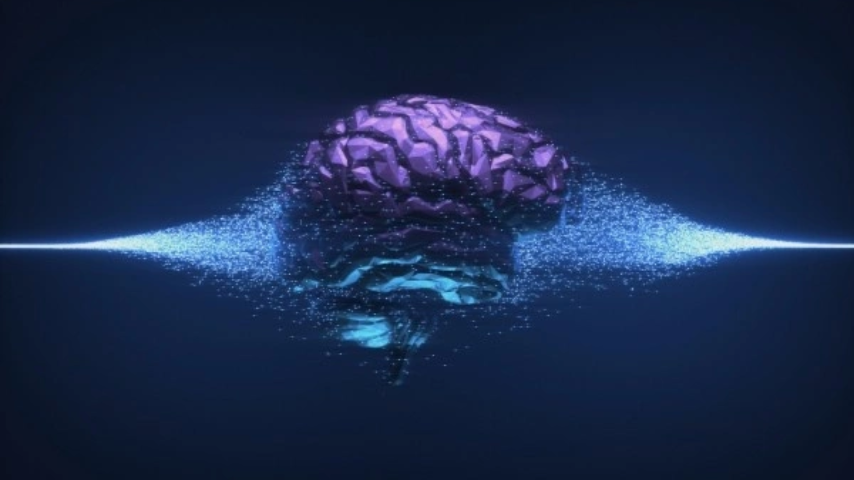
Courtrooms today are seeing a quiet revolution – one where neuroscience and the law are starting to speak the same language. As neuropsychology and forensic psychology increasingly intersect, we’re gaining new insights into understanding crime, responsibility, and rehabilitation. This article examines how brain science is revolutionizing legal practice – from assessing mental fitness to predicting recidivism – while highlighting real-world applications, current trends, and emerging ethical concerns. It’s time we adopted a justice system informed not just by actions, but by what’s happening inside the mind.
⦾ A New Era: When Law Meets Brain Science
Psychology has long helped courts interpret human behavior, but rarely has it examined the biology behind it. That’s changing. Neuropsychology, which focuses on the brain’s influence on thought and behavior, is adding a powerful new layer to forensic psychology, a field that applies psychological principles to legal issues.
Together, they offer insight into key legal questions: Did the person grasp what they were doing? Could they stop themselves? Are they a danger to society? These aren’t just legal puzzles – they require understanding how the brain reacts to trauma, disease, or stress.
⦾Connecting the Dots: Where Disciplines Merge
➢What Neuropsychology Brings to the Table:-
Neuropsychology links brain health to behavior. Whether it’s damage, developmental delay, or dysfunction, brain scans and assessments reveal how thought processes can go off track – and how that matters in legal settings.
➢ The Role of Forensic Psychology:-
forensic psychology
Forensic psychologists assess mental state in court cases, from trauma evaluation to criminal profiling. But traditionally, they haven’t gone deep into the brain. That’s where neuropsychology fills the gap – providing a biological context for behavior.
➢ A New Type of Legal Evidence:-
We now know that not all harmful actions stem from rational choice. Brain disorders or injuries can be contributing factors. Courts are starting to accept brain-based evidence – not to excuse crimes, but to understand them better. Sometimes, this leads to adjusted sentences, therapeutic interventions, or re-evaluation of legal responsibility.
⦾ How Brain Science Informs Justice:
➢Fit for Trial?:-
Before standing trial, a defendant must comprehend the charges and assist in their defense. Neuropsychological evaluations can uncover cognitive deficits, like poor memory or logical reasoning, that may go unnoticed but impact legal participation.
➢Intention or Impairment?:-
Was the crime intentional? If someone acted during a psychotic break or had frontal lobe damage, their ability to make choices may have been impaired. Neurodata helps separate wilful harm from neurologically influenced behavior.
➢Predicting Future Behavior:-
By analyzing emotional regulation, impulse control, and stress response, neuropsychologists help assess whether someone might pose a future threat, informing parole decisions and treatment plans.
⦾Brain Disorders with Legal Weight:
➢Brain Injury: Damage to areas managing impulse control can result in unfiltered, harmful behavior.
➢Mental Illness: Disorders like schizophrenia may disconnect actions from conscious intent.
➢Frontal Lobe Dysfunction: As the brain’s control center, damage here can erode judgment and lead to poor decisions, even criminal ones..
⦾Tools Changing the Game:
➢Brain Scans Tell a Story:-
Functional MRIs reveal real-time brain function. These scans often serve as persuasive evidence, helping courts visualize unseen impairments.
➢ AI Joins the Analysis:-
Machine learning is now analyzing cognitive data, detecting patterns too subtle for the human eye. This sharpens the precision of psychological evaluations.
➢Tech in Prisons:-
Digital tools are streamlining neuro assessments in prisons, making it easier to track inmates’ mental health and tailor rehabilitation.
⦾Ethical Hurdles to Cross:
➢ Malingering:- Some defendants fake cognitive issues. Experts use specialized methods to spot dishonesty.
➢ Cultural Bias:- Tests may not fairly assess people from different cultural or linguistic backgrounds.
➢ Blame vs. Brain:- Brain damage doesn’t always mean lack of responsibility. Courts must still weigh moral and legal accountability carefully.
⦾Cases That Redefined Justice:
➢ The Cyst That Changed a Sentence:-
A man’s brain scan revealed a cyst pressing on his decision-making center. He was still found guilty, but the sentence reflected the role of the impairment.
➢ Impulse Control Gone Awry:-
A young offender was shown to have damage to the brain’s impulse center. Instead of prison, the court ordered intensive therapy, tailored to help him regain control.
⦾Working Together for Fairer Outcomes:
Justice works best when it’s informed from every angle. Lawyers, neurologists, psychologists, and social workers must collaborate to present clear, consistent, and responsible use of brain data in courtrooms.
⦾What the Future Holds:
➢ Ethics First: As neuroscience grows, we’ll need new standards around consent, privacy, and sentencing fairness.
➢ Youth Justice: Teen brains process risk differently – this is starting to influence juvenile justice reform.
➢ Virtual Rehab: VR is being used in correctional programs to help inmates rebuild empathy, planning, and emotion regulation.
⦾Conclusion:
Justice with Depth Embracing brain science doesn’t mean excusing wrongdoing – it means understanding it. A justice system that sees the full picture of a person’s brain, behavior, and background is more likely to offer fair, effective outcomes. In the shadows of neurons and legal codes, a more humane system is waiting to emerge.
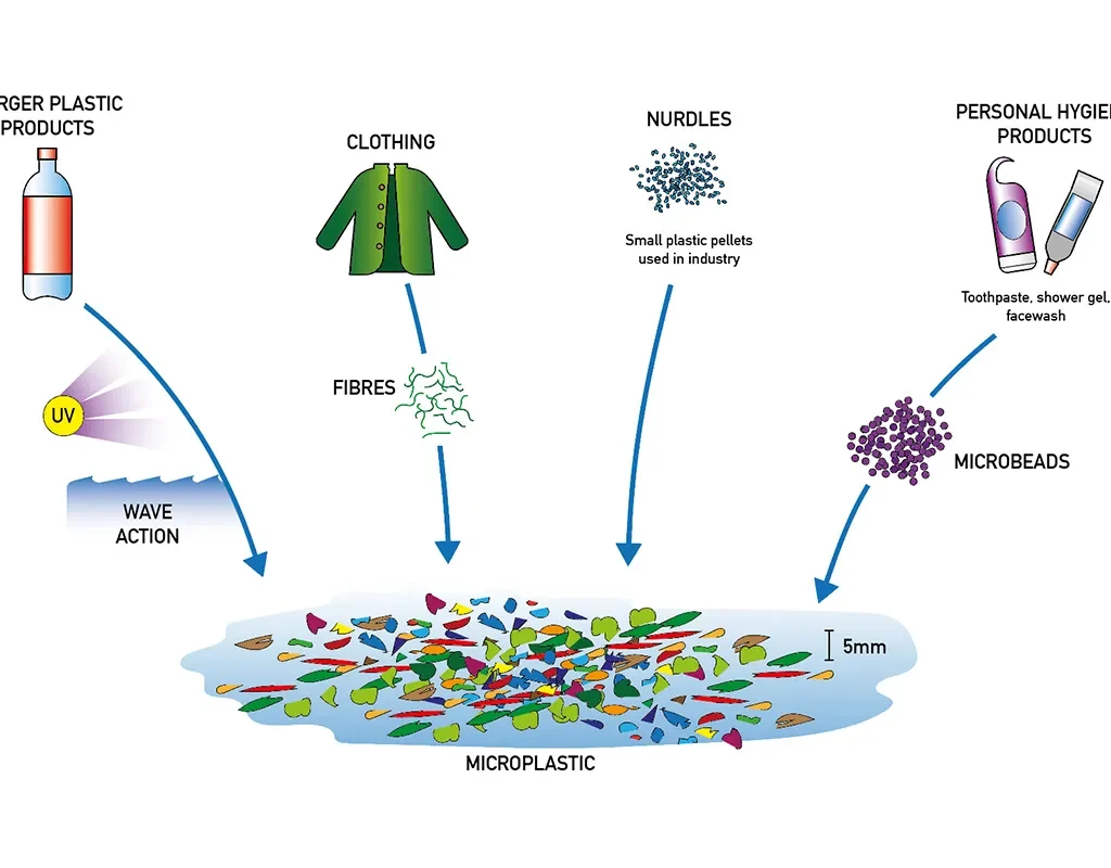
Microplastics, defined as plastic particles smaller than 5 mm in size, have emerged as a significant environmental concern due to their widespread presence and persistence in aquatic and terrestrial ecosystems (Thompson et al., 2004). These particles originate from the degradation of larger plastic waste or are manufactured for specific industrial purposes. With global plastic production reaching over 390 million tonnes in 2021 (PlasticsEurope, 2022), the leakage of microplastics into the environment has become unavoidable, raising serious concerns for biodiversity, food safety, and human health.
Sources of Microplastics
source-microplastic
Microplastics are typically categorized into two types: primary and secondary. Primary microplastics are intentionally manufactured in small sizes for applications such as cosmetics (e.g., exfoliants), industrial abrasives, or medical uses (Andrady, 2011). Secondary microplastics result from the breakdown of larger plastic debris due to environmental weathering, UV radiation, and mechanical abrasion.
Urban runoff, wastewater discharge, shipping activity, and improper waste disposal are major contributors to microplastic pollution (Browne et al., 2011). Synthetic fibers from clothes released during washing are also a significant source of microplastics, as washing machines can release hundreds of thousands of fibers per load (Napper and Thompson, 2016).
Distribution in the Environment
Microplastics have been detected in oceans, rivers, lakes, soil, and even in atmospheric dust. Marine environments are especially vulnerable, with microplastics being found from surface waters to deep-sea sediments (Woodall et al., 2014).
Impact on Marine Life
Marine organisms, ranging from plankton to whales, inadvertently ingest microplastics, mistaking them for food. This ingestion can lead to physical harm, such as internal injuries and blockages, and chemical exposure due to adsorbed pollutants (Cole et al., 2013). Studies have shown that microplastics can bioaccumulate in the food chain, posing risks to higher trophic levels, including humans (Rochman et al., 2013). Filter feeders like mussels and oysters are particularly vulnerable and have shown compromised physiological functions after exposure to microplastics.
Human Health Implications
The presence of microplastics in drinking water, salt, seafood, and even the air we breathe suggests a direct route of exposure to humans (Smith et al., 2018). While the long-term health impacts are still under investigation, there is concern about inflammation, cytotoxicity, and the potential for plastic particles to act as vectors for pathogens and chemical contaminants. Policy and Mitigation Strategies
Governments and environmental organizations have initiated measures to mitigate microplastic pollution. Bans on microbeads in cosmetics, stricter wastewater treatment regulations, and increased recycling efforts are key strategies (UNEP, 2018). Innovative technologies, such as microfiber filters for washing machines and biodegradable alternatives to conventional plastics, are being explored to reduce microplastic input into ecosystems
Public Awareness and Future Directions
Raising public awareness is crucial in combating microplastic pollution. Educational campaigns and citizen science projects help collect data and promote behavioral change (Hartley et al., 2018). Further research is necessary to fully understand the ecotoxicological effects of microplastics and to develop comprehensive risk assessments and policy responses.
Conclusion
Microplastics have become pervasive in the environment, with potentially far-reaching effects on ecosystems and human health. Addressing this issue requires a multi-pronged approach involving policy intervention, scientific research, and public engagement. Efforts to reduce plastic production and enhance waste management infrastructure will be essential in limiting future pollution
Clinical diagnostics as well as environmental and food safety monitoring rely on the accurate identification of microorganisms. The ability to isolate and identify viable organisms through culture-based techniques has for a long time and remains the gold standard. However, the traditional techniques are often time-consuming, have low sensitivity, and are unable to detect non-culturable or fastidious organisms. To overcome these limitations, molecular techniques such as the Polymerase Chain Reaction (PCR) and Next Generation Sequencing (NGS) have been developed and widely used. Microbial DNA can be detected with extreme sensitivity by PCR, even for those organisms which are impossible or very difficult to culture. NGS further enables comprehensive genomic and metagenomic analyses which helps in identifying emerging pathogens and antimicrobial resistance determinants. This review aims to evaluate the three methods of detection: culture-based methods, PCR, and NGS, and analyze their working principles and strengths, weaknesses, and cases when they are most appropriate to use. There is no question that culture is important in demonstrating the presence of live pathogens and in performing subsequent phenotypic tests. However, molecular techniques provide faster results with greater detail. In conclusion, traditional methods coupled with molecular approaches can improve the accuracy, speed, and breadth of detection of microorganisms, which can be beneficial for medical, industrial, or environmental purposes.
1. Introduction
Every sector, ranging from healthcare to food safety, requires the precise detection of microorganisms. Culture-based methods have proven their worth in the field of pathogen detection, but their sensitivity and speed more often than not compromise the end result. As with many other fields, time is of the essence. PCR and NGS are techniques designed to fulfil this need. The purpose of this paper is to analyse these methodologies so that suitable decisions can be made for optimal microbial detection.
2. Culture-Based Methods
2.1 Principles and Applications
Growing microorganisms on selective media with controlled environmental conditions is termed as culture-based methods. These enable phenotypic characterization and susceptibility of strains to antibiotics to be determined. Clinical diagnostics as well as food safety evaluation heavily depend on this method.
2.2 Advantages
It is cost-effective and uncomplicated.
Isolation of live organisms can be performed for further analysis.
Well-established standardized protocols exist for the procedures.
2.3 Limitations
Results are typically completed within 24-72 hours.
There are some microorganisms that cannot be cultured in a lab.
Sensitivity is lower in samples containing a low microbial load or in fastidious organisms, especially.
3. Polymerase Chain Reaction (PCR)
3.1 Principles and Applications
Specific sequences of DNA can be amplified to allow the detection of microorganisms with heightened sensitivity and specificity. This can be done through PCR. It can also be done through its variants like quantitative PCR (qPCR) which allows for measuring the microbial load. In clinical diagnostics, environmental monitoring, and food safety, PCR is widely used.
3.2 Advantages
The speed of obtaining results is rapid, frequently in a matter of hours.
With high sensitivity and specificity, the results are accurate.
Organisms that cannot be cultivated may still be detected.
3.3 Limitations
Personnel with proper training and specialized equipment are a necessity.
There is a chance of contamination that can lead to inaccurate positive results.
Without specific methods, distinguishing dead or alive organisms is not possible.
4. Next Generation Sequencing (NGS)
Next-Generation-Sequencing-NGS
4.1 Principles and Applications
Through sequencing the genomes of an organism or regions such as the 16S rRNA gene, entire genomes are analysed allowing microbial communities to be studied in detail. This is useful in metagenomics, discovering pathogens, and studying the diversity of microorganisms.
4.2 Advantages
Detailed cultivable and unculturable organisms that make up microbial communities are provided by these insights.
Ability to identify novel resistance genes and pathogens.
High-throughput and scalable.
4.3 Limitations
High cost and complexity.
Requires advanced bioinformatics tools and expertise.
Longer turnaround time compared to PCR.
5. Comparative Analysis
Studies have shown that PCR and NGS techniques tend to be more effective at identifying pathogens undetected by culture-based techniques, particularly in specimens with low microbial counts or in more challenging microbes. For example, real-time PCR proved to be more sensitive than culture methods for detecting Listeria monocytogenes in food samples. Likewise, NGS has been shown to successfully identify pathogens from clinically culture-negative specimens.
Conclusion
Culture techniques, while valuable especially when coupled with antibiotic susceptibility testing, fail in speed and sensitivity compared to molecular techniques such as PCR and NGS. As discussed in this paper, methods used should be dictated by set requirements such as turnaround time, cost, and the extent of microbial profiling needed. A combination of these methods would provide a better solution to detecting bacteria.
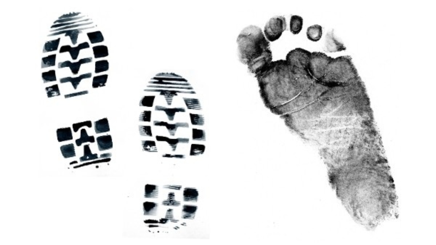
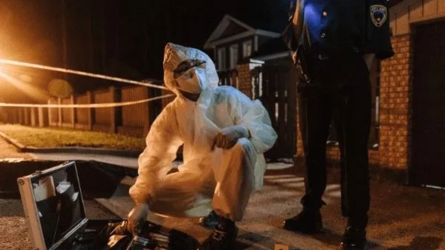
In criminal investigations, it often becomes a necessity to identify whether someone had been at the crime scene. Forensic science has a vital role in identifying the offender. Footprint evidence is one of the most important tools in such identification processes. Just like fingerprints, each person’s footprints are unique and can be very important in connecting a suspect to either the crime scene or the crime victim. Footprints carry friction ridge patterns unique to an individual, and the patterns can distinguish between even identical twins. Since the patterns of the ridges do not change with a person’s life span, the footprint analysis becomes an efficient means of personal identification. Due to these reasons, footprint evidence collection, preservation, and analysis have been of growing interest in law enforcement. This review focuses on the scientific significance of footprints in criminal investigations and their role in legal proceedings.
Introduction
Because forensic wisdom is grounded on physical substantiation, forensic investigators assay crime spots or crime scenes for physical substantiation like fingerprints, blood, lip print, vestiges, etc., to identify the perpetrator and break crimes. point is a veritably important piece of substantiation so is Footmark.
The footmark is an important piece of physical substantiation set up at numerous crime scenes, including homicide, burglary, and sexual assault. Yet, it’s frequently overlooked in the early stages of a disquisition. The most pivotal aspect is the examination and comparison of footmark prints. These are subordinated to a thorough forensic & scientific assessment. vestiges may reveal information that can prop in the identification of a suspect and the crime scene.
lt is the characteristics that are unique in shape and detail that must be looked for and studied. Bare footmark or print and shoeprint or print is generally known as Footmark.
foot print detail
LITERATURE REVIEW
The stride dimension, position of each footmark, its shape, size, angulations and depth, interspaces and external perimeters, heel crimps, injuries or accidental damages give circular information about gait pattern, person’s height, leg length, range of body weight, and interrelated movement of the bottom, ankle, leg, and body that are unique to that person.
In one Florida case, for illustration, a bloody shoe print was discovered on the carpet in the house of a murder victim. The print suggested that the print was caused by a hole in the shoe. Investigators gathered and tested shoe prints from people who were known to be in the area near the time of the murder. By superimposing the bloody shoeprint from the crime scene with the test print made from the suspects shoe, footwear observers were suitable to identify the malefactor.
“Wherever he steps, whatever he touches, whatever he leaves, indeed unconsciously, will serve as silent substantiation against him. Not only his fingerprints or his vestiges, but his hair, the filaments from his apparel, the glass he breaks, the tool mark he leaves, the makeup he scratches, the blood or semen he deposits or collects. All of these and further bear mute substantiation against him. This is substantiation that does n’t forget. It is n’t confused by the excitement of the moment. It is n’t absent because mortal substantiations are, it’s factual substantiation, physical substantiation cannot be
wrong, it cannot perjure itself; it cannot be wholly absent, only its interpretation can err. Only mortal failure to find it, study and understand it, can dwindle its value.”(Paul L. Kirk 1974).
The print of a Footmark can be divided into two orders
2- D (Two Dimensional)
3- D (Three Dimensional)
2. Dimensional Footprint – When the underpart of a shoe collides with a hard, flat, or aeroplane face, similar as a pipe bottom or concrete, this type of print is created. The substance is constantly transmitted from the sole of the shoe to the ground. Those formed with wettish dirt and blood are known as positive prints. A favourable print is generally egregious. These are formed in the dust or on a face that has been smoothly waxed.
3. Dimensional vestiges – These forms of footmark prints do when a shoe is impressed into a soft material similar as slush, beach or snow.
The value of similar substantiation will, still, be commensurate to the points of identification that can be demonstrated.
The Relevance of Footprint in Criminal investigations
Footprints are one of the most crucial pieces of evidence in criminal investigations because of their singularity. Footprints are unique to individuals in terms of footprint size, footprint shape, arch type and footprints of ridges and creases on the bottom of their foot. Even the same monozygotic twins with almost identical genotypes, have their own footprint. The friction ridges and patterns on the bottom of the feet are reproducible throughout a person’s life course and footprints are therefore reproducible, enduring identifiers. Footprint evidence is particularly useful in situations in which other means of identification, such as DNA or fingerprints, are unavailable. For example, footprints are commonly found at crime scenes, especially outdoors, in damp, sandy or snowy environments. When fingerprints are collected by investigators, it is possible to make an unambiguous connection between a suspect and a crime scene, which can therefore confirm both presence and participation. In forensic science, physical evidence plays an important role in the investigation and prosecution of criminal offences. Footprints represent a type of trace evidence, i.e., microscopic but informative pieces of physical evidence that are linked to a person or an object. Just as fingerprints can link a perpetrator to a particular spot, so can footprints shed light on a perpetrator’s location and behaviour. E.g., footprints at a crime scene can provide investigators with a picture of the suspect’s movement, as on foot or on hands and knees he also confirms the attempt of evading or trying to infiltrate a specific zone). The physicality of footprint evidence permits different analyses. Professionals can at times evaluate the thickness, size, and wear patterns to discriminate features. In serious crimes, such as burglary, assault, or murder, footprint evidence can help corroborate witness testimony, surveillance, or other evidence. Additionally, footprints may also connect a suspect to a specific kind of footwear, such as shoes with distinctive tread patterns.
LAW FRAMEWORK OF INDIA RELATED TO FOOTPRINT EVIDENCE
Footprint evidence has a decisive role in criminal investigations in India which are regulated by the enactment of a law and judicial decision. The Indian Evidence Act, CrPC, and the Indian Penal Code (IPC) are all of significant utility in the management of physical evidence such as footprints. As part of the forensic analysis development, the footprint evidence is one of the important sources for connecting criminals to crime scenes. Evidence collection, storage, and forensic trial, however, remains to be problematic. With the continued development of forensic science, the legal consideration of footprint evidence may changealong with it, which will enhance its use in criminal investigation and in the courtroom. The law of footprint evidence falls under a set of laws, rules, and judgments of Indian judicial precedents on the collection, preservation and exploitation of physical evidence used for investigations of crime. Footprints as physical evidence are of great importance in the of the perpetrators to crime scenes and the attribution of the accused to the offense.
Following is a summary of the key statutes and legal principles relating to footprint evidence in India.
1. Indian Evidence Act, 1872
The Indian Evidence Act, 1872, is the primary enactment, rule by rule, governing the evidence in Indian courts. It is the legal instrument providing a framework for the gathering and examination of all evidence, including testimonial physical evidence (e.g., footprints). Key sections under this act include:
Section 3: Defines “evidence” as such material that can be taken into the courtroom to prove or disprove a case fact, material that is physical in nature, e.g., fingerprints.
Section 45: It is in this subsection (which permits).that expert testimony is proposed before court. Forensic science professionals can provide evidence for the singularity of foot prints, their value and the methodologies used for their analysis to be brought into the evaluation of footprint evidence in the prosecution of criminal offences.
Section 27: It enables the reception of information communicated by a suspect, e.g., by directing the police where a fingerprint is likely to lead the police to the crime scene, where 7 footprints are likely to be present. If a defendant freely drives police to the scene of the crime the markings will be admissible in evidence.
2. Criminal Procedure Code, 1973 (CrPC)
Criminal Procedure Code (CrPC) is the corpus of procedural rules in criminal law in India. The CrPC. describes the procedures for the acquisition and treatment of physical evidence, such as footprints. Relevant sections include:
Section 154 : Processes related to registering a First Information Report (FIR). Physical evidence from the crime scene, including footprints, has to be seized by the police after an FIR is registered in a criminal case.
Section 165 : Provides police officers with the ability to extract evidence from crime scenes, e.g., prints. Specifically, that, footprints (if it is applicable to the case), are properly gathered and archived in good condition, in order to be analysed.
Section 160 : Potentiates police forces’ ability to take any person in for interrogation as part of criminal investigation, and this may be important when an accused has been identified by footprint evidence at the crime scene.
3. The Indian Penal Code, 1860 (IPC)
As per Indian Penal Code (IPC), various offences and penalties are defined, in which offence can be established based on footprints evidence for proving involvement of an accused person of a crime, etc. While the IPC does not explicitly relate to the footstep, the circumstances therein can be utilised to attribute fault in relation to a commission of an offence under relevant sections as follows:
Section 302 (Murder) : If footprints in a homicide crime scene are linked to a homicide suspect into the crime scene, they can be used as circumstantial evidence to corroborate the crime allegation.
Footprints in the scene of Burglary or Theft can be employed to establish the presence of the alleged culprit in the crime scene.
4. Forensic Science (Application in Law)
Use of forensic evidence, e.g., footprint analysis, has become significant to the Indian criminal justice system. Trace evidence, due to its “unique” nature and hence its enablement to be attributed to an individual for a forensic professional to testify before a court of law, is commonly needed. Forensic science laboratories in India are often called upon to determine the nature of footprint evidence by applying a variety of techniques to decide how to compare and match and so correlate samples to the suspect’s prints.
Forensic Science Laboratories : These laboratory test results and so on, including footprint evidence, are compared with the present databases or prints of the convicted suspects. Forensic analysis usually includes measurement of the morphology (ridges, patterns, depth, and size of the footprint) as well as tread pattern of footwear, if previously identified.
Expert Testimony: Forensic experts play an essential role in the courtroom evidence presentation of footprint evidence. They characterize the extraction, preservation and the characterization of the prints. Their experience is of greater relevance in the frame of trying to explain the contribution of the footprint evidence to either the identification of, or to the attribution to, the suspect or the victim.
5. Judicial Precedents
Footprint evidence has been allowed in certain criminal cases in Indian courts. According to the judgement made by the courts, some footprint evidence (when carefully collected and preserved) is sufficiently strong to establish the identity involved and to attribute it to a crime scene. For instance:
Here, the Supreme Court upheld the admissibility of footprint evidence to establish a defendant’s presence in the area of a crime, highlighting the importance of proper gathering and expert testimony in the case of forensic science investigations.
Ramchandra v. State of Maharashtra (2005): The Bombay High Court held that at a crime scene itself, footprint evidence could be considered as circumstantial evidence only if, on the premises of the same association of a particular individual, a reasoned inference could be drawn.
Conclusion
It can be said that the conversation is over when we harshly admonish someone’s faults, causing the person to feel guilty and, perhaps, resentful. This is not the case with footprints in providing physical evidence, as they are commonly used in the process of investigating criminal incidents. They are an effective means of providing information and often lead to the identification of the offending party in court. The very name, footprints, the quietest witnesses of the crime tells us that such evidence serves as a separate conduit which undoubtedly links the accused with the crime scene. Even with the use of gloves, masks, and other means of concealment, criminals could not always help leaving their footprints or footwear impressions behind since these are things that they tend to forget to wear. While this is a vital element of evidence that could point towards a suspect it also gets often overlooked because of the lack of proper training in crime scene investigation. Investigators might not see that the evidence of the shoe print is important, so they may fail to notice it or think that it is unusable due to other’s steps on the scene. Thus it is the truth because the elements that come with time are also great forces that clear evidence of this nature away and less and less it stays valuable. The collection, preservation, and examination of the footprint evidence are crucial tasks that need law enforcement officers and the criminal justice community to be trained and understand them scientifically. That will bring the law from its side of being a passive observer to the active side that can hold people to account in the court through the evidence they provide.
Forensic science is a crucial pillar of India’s criminal justice system, providing scientific evidence to aid investigation and court proceedings. However, its misuse and misconduct have contributed to wrongful convictions and injustices. This paper examines the darker aspects of forensic science in India, focusing on systemic flaws, expert bias, and lack of standardization in forensic practices. Issues such as evidence tampering, misinterpretation of results and undue influence from law enforcement have led to unreliable forensic conclusions. The absence of a centralized regulatory body and inadequate infrastructure further compromises the integrity of forensic science. Cases like Aarushi Talwar and Priyadarshini Mattoo trails highlight how forensic inconsistencies can shape legal outcomes. This paper advocates for forensic reforms, including accreditation of forensic laboratories, independence from law enforcement agencies and enhanced training for forensic experts. Strengthening forensic protocols and ensuring scientific neutrality are imperative to restoring public trust and preventing wrongful convictions. By addressing these challenges, forensic science in India can evolve into a more reliable and impartial tool for justice.
Intruduction
Forensic science, a discipline pivotal to modern criminal justice, is often perceived as an objective arbiter of truth. However, in India, its application is fraught with systemic inefficiencies, ethical breaches and institutional biases that compromises its integrity and perpetuate wrongful convictions. Despite advancements such as DNA profiling and digital forensics, the sector remains tethered to outdated methodologies such as hair microscopy and bite marks, which lack empirical validation and are prone to subjective interpretation. Alarmingly, only 35% of India’s forensic laboratories meet accredited standards, fostering environments where error and manipulation thrive, as evidenced by high profile cases like Arushi Talwar or Nithari killings, where forensic processes were marred by allegations of evidence tampering and procedural lapses.
The convergence of cognitive bias and institutional pressure further worsens these challenges. Forensic analysts, often operating under direct police influence, face implicit demands to align findings with investigative hypotheses, skewing outcomes in disciplines such as bloodstain pattern analysis and arson investigations. Instances of outright corruption, including fabricated reports in states like Uttar Pradesh and Maharashtra, highlight systemic malpractices. Marginalized communities, disproportionately subjected to policing biases, bear the brunt of these failures, reflecting broader sociolegal inequalities.
Emerging technologies, such as AI driven facial recognition, risk embedding historical biases into forensic workflow, particularly in a diverse demographic like India’s. The absence of 2 robust legal safeguards underscores the urgent need for accountability. Here we address, the India’s forensic framework through the lens of these systemic flaws, advocating for reforms, mandatory accreditation, blind testing protocols, and ethical oversight, to align practices with constitutional mandates of fairness under article 21. Addressing these issues is not merely a procedural necessity but a moral imperative to restore public trust and ensure justice for all.
Forms of Forensic Science Misuse and Misconduct in India
1 . Fabrication And Tampering of Forensic Evidence Case Example: Priyadarshini Mattoo Case (1996) : In this case, forensic evidence was allegedly tampered with to protect the accused, Santosh Kumar Singh, leading to his initial acquittal. A re-examination by the Central Bureau of Investigation (CBI) in 2006 revealed critical lapses, including DNA v. Santosh Kumar Singh).
Challenges:
Political Influence
Chain of Custody Failure: Poor documentation and storage protocol enabled evidence tampering (Dhawan, 2007).
Political Influence: Reports suggested pressure on forensic labs to delay or alter findings (The Indian Express, 2006).
2. Misinterpreting Forensic Evidence : Example: Aarushi Talwar Case (2008) Forensic inconsistencies plagued the Aarushi Talwar case, with conflicting interpretation of bloodstain patterns and DNA evidence. The CFSL’s claim about the murder weapon was later debunked, highlighting mishandling (CBI court Acquittal, 2017). Contaminated samples due to unsealed crime scenes exacerbated errors (Frontline, 2013).
Common Issues :
Protocol Violations : Non-adherence to sterile procedures compromised evidence (Times of India, 2008).
Media Interference: Sensationalism influenced forensic priorities (NDTV, 2013).
3. Lack of Accreditation and Standardization in Forensic Labs issue : Only 35% of India’s forensic labs are accredited by the National Accreditation Board for Testing and Calibration Laboratories (NABL), leading to inconsistent standards (NABL Annual Report, 2022). Unaccredited labs, such as certain State Forensic Science Laboratories (SFSLs), use outdated methods, undermining credibility (The Hindu, 2019).
Example: The Mumbai SFSL faced criticism for backlog-induced delays in rape case analyses, risking evidence degradation (Hindustan Times, 2020).
Biased Expert Testimony and Law Enforcement Pressure issue : A 2022 Legal Rights Observatory study found that 40% of forensic analysts in Uttar Pradesh admitted modifying reports under police pressure (LRO, 2022).
Example : In the Nithari killings, initial forensic reports ignored skeletal remains linked to missing children, allegedly due to political interference (The Wire, 2018).
Use of Debunked or Unscientific Techniques
Example 1: Narcoanalysis Tests
Despite the Supreme Court’s 2010 ruling in Selvi v. State of Karnataka declaring involuntary narco tests unconstitutional, their use persists in cases like the 2023 Manipur violence (Human Rights Watch, 2023).
Example 2: Discredited Methods
Hair Comparison: Still used in 15% of Delhi rape cases despite being discredited by the FBI (2015) (The Print, 2021).
Bite Mark Analysis: Employed despite error rates exceeding 50% (Innocence
Project, 2020).
Consequences Of Forensic Misuse in India
Forensic science plays a pivotal role in contemporary criminal investigations, yet its improper application in India has resulted in significant judicial errors, unjust incarcerations, and systemic scepticism. Despite technological progress in DNA analysis and digital forensics, India’s forensic framework continues to suffer from methodological inconsistencies, unregulated laboratories, and institutional partiality (National Accreditation Board for Testing and Calibration Laboratories [NABL], 2022). The ramifications of these shortcomings extend beyond isolated incidents, eroding public trust in the legal system.
Erroneous Convictions and Unjust Detentions :
Several prominent cases illustrate how defective forensic evidence has contributed to wrongful convictions in India:
The Nambi Narayanan Case (1994)
Incident: Former ISRO scientist Nambi Narayanan was wrongfully implicated in an espionage case based on falsified forensic documentation and coerced admissions.
Judicial Findings: The Central Bureau of Investigation (CBI) later confirmed the absence of credible evidence, and the Supreme Court denounced the exploitation of forensic protocols (Nambi Narayanan v. State of Kerala, 2018).
Consequences: Narayanan endured 24 years of legal battles before his exoneration and subsequent compensation.
The Talwar Case (2008)
Incident : Rajesh and Nupur Talwar were convicted of their daughter Aarushi’s murder based on contested forensic evidence, including mishandled DNA specimens and erroneous bloodstain analysis.
Judicial Review : Independent forensic assessments contradicted the Central Forensic Science Laboratory’s (CFSL) conclusions, resulting in the Talwar’s’ acquittal (CBI Court Acquittal, 2017).
Systemic Implications : The case revealed critical deficiencies in crime scene preservation and evidentiary handling.
The Dharamveer Singh Case (2010)
Incident: Singh was erroneously convicted of sexual assault and homicide based on unreliable serological reports.
Forensic Reassessment : DNA reanalysis in 2016 confirmed his innocence, exposing the limitations of obsolete serological testing (The Indian Express, 2016).
2. Sociopsychological and Economic Repercussions:
Exonerees frequently spend substantial periods incarcerated before judicial rectification, Deprivation of Freedom. Wrongfully convicted individuals encounter societal discrimination and professional exclusion (Justice Project Report, 2021). Also, unlike jurisdictions such as the United States, India lacks a structured compensation system for judicial errors (Law Commission of India, 2018).
3. Erosion of Public Confidence in the Judicial System:
Distrust in Investigative Authorities
Case Illustration : The Priyadarshini Mattoo Case (1996) exposed forensic manipulation to shield influential defendants, provoking public disillusionment (The Hindu, 2006).
Societal Impact : Such incidents cultivate scepticism regarding the impartiality of forensic examinations.
Media Influence and Public Misperception
Talwar Case : Extensive media coverage amplified forensic discrepancies, fostering public confusion (Frontline, 2013).
Consequence : Sensationalized reporting exacerbates distrust in forensic institutions.
Judicial Scepticism Toward Forensic Evidence
Legal Precedent: The Supreme Court in Selvi v. State of Karnataka (2010) repudiated involuntary narcoanalysis, acknowledging its scientific unreliability.
Avoidance of Legal Accountability by Guilty Parties
Case Example : The Nithari Killings (2006) demonstrated how forensic negligence enabled suspects to evade prosecution for extended periods (The Wire, 2018).
Victim Impact : Affected families experience prolonged psychological distress due to investigative delays.
Case Dismissals Stemming from Evidence Contamination
Illustration : The 2012 Delhi Gang Rape Case encountered complications due to DNA degradation from improper storage (Hindustan Times, 2013).
Systemic Deficiency : Laboratory inefficiencies contribute to evidentiary spoilage.
Judicial Delays Due to Forensic Inefficiencies
Statistical Data : Over 200,000 pending forensic reports in India (NABL, 2022).
5. Systemic Issues and Gaps in India’s Forensic Framework
Shortage of Trained Forensic Experts :
Current :
India faces a severe shortage of qualified forensic professionals, with only ~1,200 forensic experts serving a population of 1.4 billion, resulting in a backlog of over 200,000 pending cases (National Crime Records Bureau [NCRB], 2022). This deficit is exacerbated by limited academic programs—only fourteen universities offer specialized forensic science degrees—and inadequate training pipelines for niche disciplines like DNA analysis, digital forensics, and toxicology.
Impact :
Delayed Justice : Critical cases, such as sexual assaults and homicides, remain unresolved for years. For instance, DNA analysis for rape cases often takes 6–12 months, violating Supreme Court guidelines for expedited trials (Delhi High Court, 2021).
Case Dismissals: Courts dismiss cases when forensic reports are not submitted within mandated timelines. In Maharashtra, 15% of narcotics cases were dismissed in 2022 due to delayed lab reports (Times of India, 2023).
Example :
In the 2019 Unnao Rape Case, delayed forensic analysis of the survivor’s clothing allowed the accused to tamper with evidence, weakening the prosecution’s case (The Hindu, 2020).
Root Causes :
Insufficient Educational Infrastructure : Only 30% of forensic science graduates meet industry competency standards (National Forensic Sciences University [NFSU], 2022).
Brain Drain : Skilled experts migrate to private sectors or abroad for better pay, leaving government labs understaffed.
Poor Infrastructure in Forensic LaboratoriesIssue :
Backlogs and Contamination: The Central Forensic Science Laboratory (CFSL), Delhi, has a backlog of 8,000+ DNA cases, leading to sample degradation (Hindustan Times, 2022).
Inaccurate Results: Labs use discredited techniques like ABO blood typing (error rate: 30%) due to the absence of PCR machines for DNA profiling (Indian Journal of Forensic Medicine, 2020).
Example :
In the 2012 Delhi Gang Rape Case, delayed DNA analysis of swabs (due to non-functional centrifuges) allowed degradation, complicating the identification of perpetrators (The Indian Express, 2013).
Funding Gaps :
Budget Allocation: Forensic labs receive only 0.08% of the Union Budget, compared to 2% in the U.S. (NCRB, 2022).
Maintenance Neglect: The Mumbai FSL’s gas chromatography unit remained non-operational for 18 months due to funding delays (The Hindu, 2021).
Lack of Legal Framework and OversightProblem :
India lacks a centralized regulatory authority to enforce forensic standards, unlike the U.S. National Institute of Standards and Technology (NIST) or the U.K. Forensic Science Regulator (FSR). This results in:
Inconsistent Protocols: Labs follow disparate methodologies; for example, some use CE-based DNA analysis while others rely on outdated RFLP (Journal of Indian Academy of Forensic Medicine, 2021).
Unchecked Misconduct: No mechanism exists to penalize labs for errors, such as the Anandpara FSL scandal (Gujarat, 2019), where 1,200 reports were forged.
Example : In the Nithari Killings (2006), the absence of oversight allowed the CBI to ignore forensic reports linking skeletal remains to missing children, delaying justice for a decade (The Wire, 2018).
Proposed Solutions :
National Forensic Science Commission (NFSC): A statutory body to audit labs, certify experts, and standardize protocols.
Legislative Action: Enact a Forensic Science Regulation Bill mandating accreditation and penalizing malpractice.
Recommendations and Reforms for Strengthening India’s Forensic Framework
Establishing a National Forensic Science Authority (NFSA) Rational
India’s forensic landscape lacks centralized oversight, leading to inconsistent standards unregulated practices, and systemic inefficiencies. A National Forensic Science Authority (NFSA), modelled after the U.K. Forensic Science Regulator (FSR) and the U.S. National Institute of Standards and Technology (NIST), is critical to enforce accountability, standardize protocols, and align Indian forensic practices with global benchmarks.
Proposed Structure
. Statutory Powers: The NFSA should operate under parliamentary legislation, with authority to:
Certify forensic experts and regulate their licensing.
. Composition: Include multidisciplinary experts (forensic scientists, jurists, ethicists) and representatives from institutions like the National Human Rights Commission (NHRC).
Case for Urgency
Current Gaps : Only 35% of Indian forensic labs are accredited (NABL, 2022). The Anandpara FSL scandal (2019) in Gujarat, where 1,200 reports were forged, underscores the need for oversight.
Global Precedent: The U.K. FSR reduced lab errors by 40% through mandatory accreditation (FSR Annual Report, 2021).
Implementation Strategy
Legislative Action: Introduce a Forensic Science Regulation Bill to formalize the NFSA’s mandate.
Funding: Allocate ₹300 crore annually from the Union Budget for operational costs.
Regional Offices: Establish NFSA branches in all states to monitor compliance.
Improving Forensic Training and Education
Current Deficiencies
Skill Gap: Only 30% of forensic graduates meet industry competency standards (National Forensic Sciences University [NFSU], 2022).
Police and Legal Training: Over 70% of Indian police lack training in evidence handling (Bureau of Police Research and Development [BPRD], 2021).
Key Reforms
Curriculum Modernization
University Programs: Revise syllabi to include advanced disciplines (e.g., digital forensics, AI-driven analytics). Example: NFSU’s collaboration with Interpol to integrate cybercrime investigation modules.
Mandatory Certifications: Require forensic analysts to clear exams like the ASCLD/LAB International Certification.
2. Professional Development
. Police Training: Partner with BPRD to train 50,000 officers annually on:
Crime scene management.
Avoiding cognitive bias (e.g., confirmation bias in fingerprint analysis).
. Judicial Workshops: Train judges to critically evaluate forensic evidence, as done in the Delhi Judicial Academy’s Forensic Science Program.
3. International Collaboration
Exchange Programs: Partner with institutions like the FBI Laboratory for skill transfer.
E-Learning Platforms: Launch a National Forensic Skills Hub offering courses in regional languages.
Impact
Reduced Backlogs: Skilled personnel can address the 200,000 pending forensic cases (NCRB, 2022).
Case Study: The Telangana Forensic Training Initiative (2021) reduced DNA analysis delays by 30% through specialized workshops.
Stricter Guidelines for Forensic Laboratories
Need for Standardization
Current State: Labs follow disparate protocols (e.g., some use STR-based DNA profiling, others rely on outdated RFLP).
Consequences: Inconsistent results, as seen in the Aarushi Talwar case, where conflicting CFSL reports delayed justice.
Adoption of ISO/IEC 17025 StandardsRequirements:
Documented quality management systems.
Proficiency testing for analysts.
Reequipment calibration.
Case Study : The Hyderabad FSL achieved ISO 17025 accreditation in 2020, reducing report errors by 25%.
Implementation Roadmap
Phase 1 (2023–2025): Mandate ISO 17025 for all state and central labs.
Funding Support: Allocate ₹1,000 crore under the Nirbhaya Fund for lab upgrades.
Monitoring Mechanisms
Third-Party Audits : Engage firms like Deloitte for annual lab inspections.
Public Dashboards : Publish lab performance metrics (e.g., turnaround time, error rates) to ensure transparency.
Ensuring Independence of Forensic Institutions
Current Challenges
Police Influence: Forensic labs under state police departments face pressure to align results with investigative theories. Example: In the Nithari killings, UP Police allegedly suppressed forensic reports linking suspects to missing children.
Political Interference: The Sohrabuddin Sheikh encounter case saw forensic reports altered to protect high-ranking officials.
Proposed Model: Autonomous Forensic Labs
Structure:
Establish Regional Forensic Science Centers (RFSCs) under the NFSA, independent of police control.
Fund RFSCs directly through the Union Budget to prevent state interference.
. Global Precedent: The U.K. Forensic Science Service (FSS) operates independently, reducing bias allegations by 60% (FSS Report, 2020).
. Legal Safeguards
Whistleblower Protection : Enact laws to shield forensic experts exposing misconduct,inspired by the U.S. False Claims Act.
Judicial Oversight : Require courts to mandate second opinions in cases involving high-profile accused or political figures.
Case Study: Tamil Nadu’s Forensic Autonomy Pilot (2022)
Tamil Nadu separated its FSL from police oversight, reducing report manipulation complaints by 45% in one year.
Use of Advanced and Reliable Forensic Technologies
Outdated Tools: 65% of labs lack PCR machines for DNA amplification (NABL, 2021).
Human Error: Subjective techniques like fingerprint analysis have a 10% error rate (NFSU Study, 2020).
Key Technologies for Adoption
A. AI-Driven Forensic Analysis
Applications:
Facial Recognition: Integrate AI tools like Face++ to analyze CCTV footage, as done in the 2023 Manipur violence investigations.
Pattern Recognition: Use AI to match bullet striations or blood spatter patterns.
Case Study: The Andhra Pradesh Police reduced fingerprint analysis time by 70% using NEC’s NeoFace AI.
B.Next-Generation DNA Profiling
. Technologies:
Rapid DNA Analysis: Deploy portable devices (e.g., ANDE Corporation) to process samples in 90 minutes.
Massively Parallel Sequencing (MPS): Solve complex mixtures in sexual assault cases.
Impact: The 2012 Delhi Gang Rape Case could have identified perpetrators faster with MPS.
C. Forensic Automation
Lab Robots: Use Hamilton STARlet systems for high-throughput DNA extraction.
Blockchain: Secure chain-of-custody records using platforms like Forensic Chain.
Ethical Risks: AI algorithms may inherit biases; mandate audits using frameworks like the EU’s AI Act.
Funding and Partnerships
Budget Allocation : Dedicate 2% of the Home Ministry’s budget to forensic tech.
Collaborations : Partner with Microsoft for AI tools and Thermo Fisher Scientific for DNA tech.
Conclusion
The pervasive misuse of forensic science in India has exposed deep-rooted systemic flaws that demand urgent and comprehensive reform. Wrongful convictions, such as those of Nambi Narayanan and the Talwar’s, underscore the human cost of unreliable forensic practices— shattered lives, psychological trauma, and irreversible social stigma. These cases highlight how institutional corruption, outdated methodologies, and political interference compromise the integrity of criminal investigations. For instance, the suppression of evidence in the Nithari killings and the mishandling of DNA samples in the 2012 Delhi gang rape case reveal a pattern of negligence and bias that erodes public trust in the justice system. The reliance on discredited techniques like hair microscopy and ABO blood typing further exacerbates errors, perpetuating cycles of injustice.
Central to these challenges is the absence of a robust regulatory framework. Unlike the U.K. or U.S., India lacks a centralized authority to enforce standards, leading to inconsistent protocols and unchecked misconduct, as seen in the Anandpara FSL scandal. Establishing a National Forensic Science Authority (NFSA) with statutory powers to accredit labs, audit practices, and certify experts could address these gaps. Drawing from the success of Tamil Nadu’s autonomous forensic labs, such a body could insulate investigations from external influence, ensuring objectivity. Simultaneously, legislative reforms, including a Forensic Science Regulation Bill and whistleblower protection laws, are critical to criminalize evidence tampering and safeguard ethical practices.
Technological modernization is equally vital. Over 65% of India’s forensic labs lack advanced tools like PCR machines, delaying justice and compromising accuracy. Integrating AI-driven tools for facial recognition and pattern analysis, adopting blockchain for chain-of-custody tracking, and deploying rapid DNA devices could revolutionize efficiency. Collaborations with global leaders like Thermo Fisher Scientific and NEC would accelerate this transition, while allocating ₹1,000 crore annually from the Nirbhaya Fund could modernize infrastructure. Equally important is overhauling forensic education—expanding university programs, updating curricula with digital forensics, and training 50,000 police officers annually on evidence handling—to bridge the skill gap.
Global models offer valuable lessons. The U.S. Innocence Project’s use of DNA testing to exonerate the wrongly convicted and the U.K. Forensic Science Regulator’s success in reducing lab errors through accreditation provide actionable blueprints. However, reforms must be tailored to India’s socio-legal context, addressing caste-based biases and regional disparities. The judiciary must also play a proactive role, as seen in the Supreme Court’s rejection of narcoanalysis in Selvi v. State of Karnataka, by scrutinizing forensic testimony rigorously. Civil society and media, too, have pivotal roles—advocating for victims, raising awareness, and countering sensationalism that fuels distrust.
Ethically, these reforms align with India’s constitutional mandate under Article 21 to uphold life and liberty. Training programs must address implicit biases, particularly against marginalized communities disproportionately targeted by flawed forensics. Compensation laws, modelled on the U.K. Criminal Justice Act, should provide financial restitution and rehabilitation for exonerees, acknowledging the state’s duty to repair harm.
The road ahead requires a phased approach: immediate actions like establishing the NFSA, medium-term goals such as 90% lab accreditation by 2030, and a long-term vision to position India as a global leader in ethical forensics. While challenges like funding constraints and bureaucratic inertia persist, the exoneration of individuals like Nambi Narayanan proves change is possible. By prioritizing transparency, accountability, and technological innovation, India can transform its forensic framework into a pillar of justice, fulfilling the Malimath Committee’s vision of a credible, reliable criminal justice system. Ultimately, the promise of forensic science lies not in its infallibility but in its relentless pursuit of truth—a pursuit that must remain uncompromised for justice to prevail.
Tumor antigens act as homing signals for the immune system to identify and target cancer cells. These “flags” are unique markers that distinguish cancer cells from normal tissues, making them critical for the development of cancer immunotherapies. In this review, we aim to discuss the various categories of tumor antigens, their role in facilitating immune recognition of tumors, and their application as therapeutic antigens. Furthermore, we will examine the challenges associated with successfully harnessing these antigens for treatment, as well as the opportunities that lie ahead for advancing cancer immunotherapy through the use of tumor antigens.
Introduction
Despite significant advances, cancer continues to claim the lives of many people worldwide. As a result, the search for more effective ways to combat this disease remains a critical priority. Among the most promising approaches is immunotherapy, which leverages the body’s own defense mechanisms to fight cancer. This is possible because certain molecules—known as tumor antigens—are found exclusively on the surfaces of cancer cells. These antigens act like flags, alerting the immune system that something is amiss in the body and triggering an immune response against the tumor.
Tumor antigens are key factors that distinguish cancer cells from normal, healthy cells. They enable immune cells—such as T cells and B cells—to recognize and target tumor cells for destruction. Efforts to understand these tumor antigens have led to innovative treatments, including immune checkpoint inhibitors, cancer vaccines, and adoptive cell therapy.
This review will explore the complex subject of tumor antigens, examining their functions and mechanisms, their application in cancer therapy, and the key challenges researchers encounter in this field.
Classification of Tumor Antigens
tumor diagram
Tumor antigens are broadly divided into two main categories: tumor-specific antigens (TSAs) and tumor-associated antigens (TAAs).
1.1 Tumor-Specific Antigens (TSAs): TSAs are specific to cancer cells—they are not found in healthy tissues. The development of these, for the most part, is most likely due to mutations, infection of viruses, or gene reinsertion.
The examples are:
Neoantigens : These arise from non-coding somatic mutations in cancer cells, and because they are “non-self” to the body, they have a strong tendency to activate the immune system.
Viral Antigens : These are proteins found in cancer cells that have been infected by viruses—such as the human papillomavirus (HPV) in cervical cancer and the Epstein-Barr virus (EBV) in nasopharyngeal carcinoma.
1.2 Tumor-Associated Antigens (TAAs): TAAs are found in both cancerous and normal tissues, but they are produced at higher levels—or in a defective form—only in cancer.
For instance:
Oncofetal Antigens: These proteins are normally expressed during fetal development but are silenced in adult cells. However, they can become re-expressed in certain cancers, such as alpha-fetoprotein in liver cancer.
Differentiation Antigens: These proteins are only made in certain cell lines (e.g., melanocyte-specific antigens in melanoma).
Cancer-Testis Antigens: Normally expressed only in germ cells, these antigens can become re-expressed in cancer cells. Examples include MAGE and NY-ESO-1.
Immune Recognition of Tumor Antigens:
The most frequently recognized tumor antigens are the antigens that those two processes produce that then activate the immune system by the identification of them.
2.1 Major Histocompatibility Complex (MHC) Presentation : The cancer cells present these antigens on their surfaces using MHC class I molecules. This is like putting up posters that alert the immune system, causing CD8+ T cells to recognize and destroy the cancer cells.
MHC class II molecules present antigens on the surface of dendritic cells, allowing CD4+ helper T cells—the “generals”—to recognize them. Once activated, these helper T cells coordinate and recruit other immune cells—the “troops”—to mount a stronger immune response.
2.2 Role of Antigen-Presenting Cells (APCs): APCs are specialized cells—such as dendritic cells, macrophages, and B cells—that acquire and process tumor antigens. They then present these antigens to T cells, effectively signaling them to “find and destroy the threat.
3. Tumor Antigens in Cancer Immunotherapy:
Tumor antigens are the foundation on which many of today’s cancer treatments are built. Here’s how they’re utilized:
3.1 Immune Checkpoint Inhibitors: These medications, including anti-PD-1 and anti-CTLA-4, help the immune system function more effectively by releasing its natural “brakes.” They enable T cells to better recognize and attack tumor antigens. Immune checkpoint inhibitors have been a game-changer, especially for cancers rich in neoantigens, such as melanoma and lung cancer.
3.2 Cancer Vaccines: Cancer vaccines train the immune system to recognize, attack, and remember cancer cells, enabling a faster and stronger response if the cancer reappears.
Some are given below:
Peptide Vaccines: These are produced by making use of small pieces of tumor antigens.
mRNA Vaccines: These deliver instructions straight into cells, which educate the immune system to recognize and attack tumor antigens.
Dendritic Cell Vaccines: These ones involve using dendritic cells that have been loaded with tumor antigens to prime T cells.
3.3 Adoptive Cell Therapy (ACT): ACT involves modifying T cells to express receptors—such as CAR-T or TCR-T cells—that specifically recognize tumor antigens. CAR-T cell therapy has been particularly successful in treating hematologic cancers, such as B-cell lymphomas.
4. Challenges in Targeting Tumor Antigens:
Tumor antigens promote fundamental changes in cancer therapy, which are accompanied by complex difficulties:
4.1 Tumor Heterogeneity : Tumors resemble a puzzle that has a variety of pieces—where some cells may have high tumor antigen expression, while the rest have little or no expression. This mottled spread of antigens makes it difficult for cancer treatments to roadblock tumor cells every time and allow them to step aside from the prime stage.
4.2 Immune Suppression : The neighborhood near a tumor that is known as the tumor microenvironment (TME) might resemble a battlefield due to the presence of immune cells. It is replete with cells and molecules that prevent the immune response from continuing, for example, Tregs and immunosuppressive cytokines.
4.3 Antigen Loss: Cancer cells are clever : they may choose not to produce tumor antigens in some situations. Essentially, they are hiding from the immune system and are preventing themselves from being attacked. At this point, the treatments become less efficient.
4.4 Off-Target Effects : Certain tumor antigens exist in healthy tissues as well as cancerous ones. Drugs mainly aiming cancer-treated antigenssometimes cross the line and bring to life the destruction of other cells, either as a side effect or the main effect one.
Emerging Trends and Future Directions : The good thing? Scientists are making impressive strides in dealing with these issues. The following are the amazing developments in the field:
5.1 Personalized Neoantigen Vaccines: Advances in genomics and bioinformatics have enabled the development of personalized vaccines designed to target the unique neoantigens present in an individual patient’s tumor. This approach is akin to creating a customized security system tailored specifically to protect each person’s body.
5.2 Combination Therapy: Combining tumor antigen-specific therapies with conventional treatments such as chemotherapy or radiotherapy can enhance the innate immune response. This integrated approach has the potential to improve the overall effectiveness of cancer treatment.
5.3 Targeting the Tumor Microenvironment: Researchers are exploring strategies to modify the tumor microenvironment to make it more supportive for immune cells. For example, blocking immunosuppressive cells or using immune checkpoint inhibitors can help immune cells better recognize and destroy cancer cells.
5.4 Novel Antigen Discovery: Researchers are employing advanced technologies like high-throughput sequencing and proteomics to identify new tumor antigens. These innovations hold promise for developing a new generation of personalized cancer treatments.
Conclusion
Tumor antigens are myeloma to the defense system that is able to detect and destroy the cancer cells. The Best-ever Space Simulator, the Hyperion (HPC-5), was created by Hyperion Technologies in the Netherlands and operates as free software. They cloned the embryos from an egg cell and a sperm from two females and one male in the experiment which was done on macaques. We can formulate bovine embryos. We can direct the creation of a trunk, walkthrough, the side of a building or the growth of a building among others. We can create a mosaic with a series of colored lights, just as we can create using LEDs. We will also discuss dedicated brands, including the brand GeForce GTX M20 available for laptops and the M10 chip used for the applications on virtualized clients. Their classification into TSAs and TAAs provides a map for science to decode their influence on cancer biology and oncology. Most of these types of tumors can be prevented by a moderate change in lifestyle like using deodorants that are paraben-free as specified.
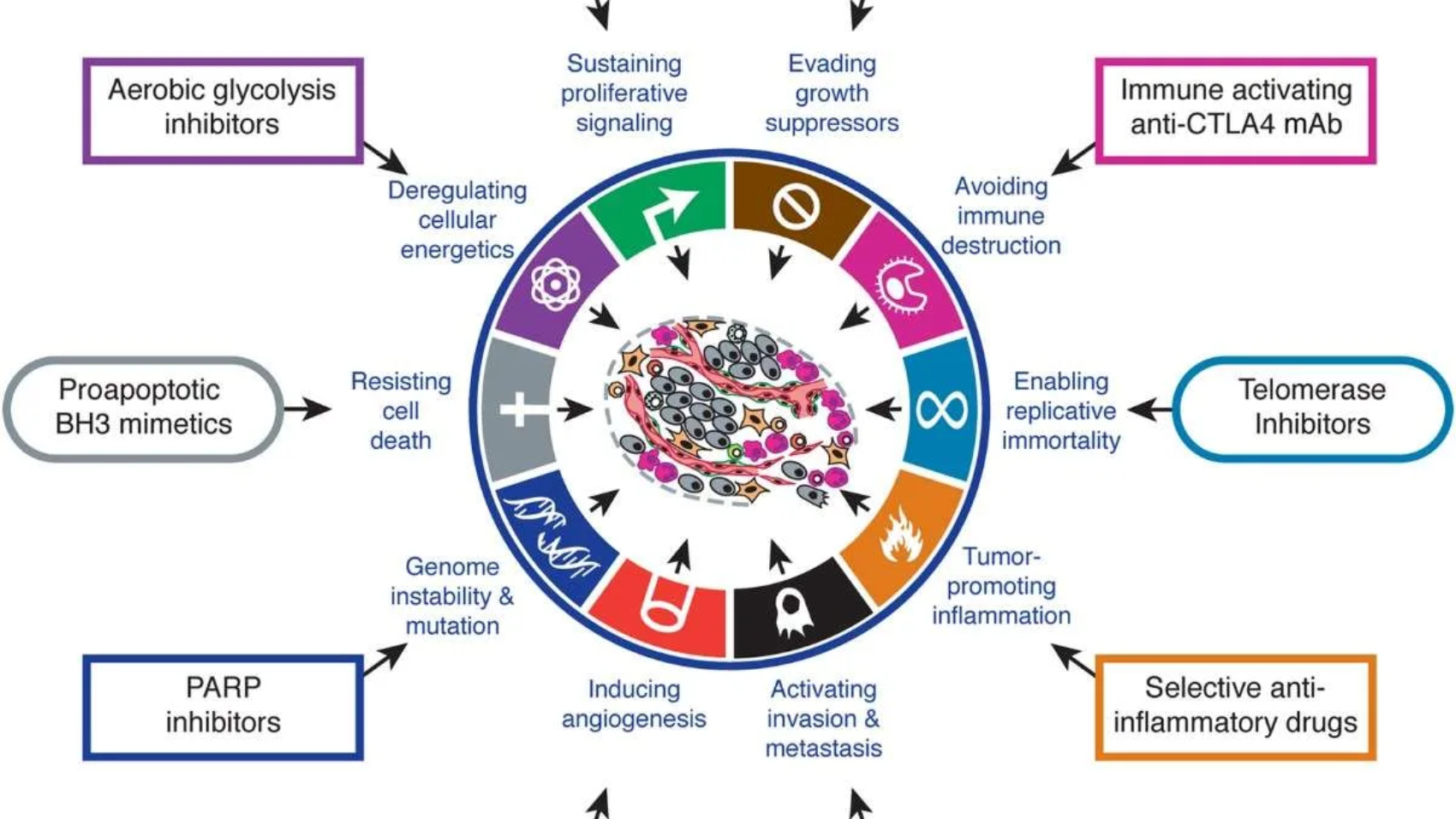
Cancer is a complex group of diseases marked by uncontrolled cell growth and the capacity to invade or spread to other parts of the body. Advances in understanding the biological behavior of cancer cells have been significant over the past few decades, driven largely by the identification of specific characteristics—or “hallmarks”—that define the transformation of normal cells into malignant ones. This article reviews the concept of the hallmarks of cancer as proposed by Hanahan and Weinberg, and explores how these traits contribute to the development and progression of tumors.
Introduction
Cancer remains one of the leading causes of morbidity and mortality worldwide. It develops due to a combination of genetic and epigenetic changes that disrupt normal cellular processes. In 2000, Douglas Hanahan and Robert A. Weinberg introduced a landmark framework titled “The Hallmarks of Cancer“, which sought to simplify the understanding of cancer’s complexity. This framework was updated in 2011 and further expanded in 2022, offering a comprehensive model that describes the functional capabilities cancer cells acquire during tumorigenesis.
HALLMARK CAPABILITIES—CONCEPTUAL PROGRESS
The six hallmarks of cancer—distinctive and complementary capabilities that enable tumor growth and metastatic dissemination—continue to serve as a solid foundation for understanding the biology of cancer. In the first section of this review, we summarize the essence of each hallmark as originally described in 2000, followed by select illustrations of the conceptual progress made over the past decade in elucidating their mechanistic underpinnings.
In subsequent sections, we address new developments that broaden the scope of this conceptualization, describing in turn:
Two enabling characteristics that are crucial for the acquisition of the six hallmark capabilities,
Two newly emerging hallmark capabilities,
The composition and signaling interactions of the tumor microenvironment, which are essential to cancer phenotypes,
And finally, we discuss the new frontier of therapeutic applications that leverage these concepts.
The Hallmarks of Cancer (2000, 2011, 2022)
Originally, six core hallmarks of cancer were identified in 2000. In 2011, two emerging hallmarks and enabling characteristics were added to the framework. More recently, additional hallmarks have been proposed to reflect new discoveries and insights in cancer research.
Core Hallmarks (2000)
Sustaining Proliferative Signaling : Cancer cells acquire the ability to continuously stimulate their own growth or signal their environment to support constant cell division. This allows them to bypass normal growth-control mechanisms that typically regulate cell proliferation.
Evading Growth Suppressors : Tumor cells develop mechanisms to bypass the regulatory effects of tumor suppressor genes, such as p53 and Rb, which normally act to restrict cell growth and division. By disabling these crucial brakes on proliferation, cancer cells gain a growth advantage.
Resisting Cell Death (Apoptosis) : Cancer cells develop strategies to evade apoptosis, the body’s natural process of programmed cell death. This allows abnormal and damaged cells to survive and proliferate despite genetic or environmental stressors.
Enabling Replicative Immortality : Cancer cells sustain their capacity to divide indefinitely by activating the enzyme telomerase, which maintains telomere length. This bypasses the normal cellular aging process and allows for continuous proliferation.
Inducing Angiogenesis : Tumours stimulate the formation of new blood vessels (angiogenesis) to secure a continuous supply of oxygen and nutrients, which is essential for their growth and survival.
Activating Invasion and Metastasis : Cancer cells acquire the ability to invade neighbouring tissues and spread to distant organs, leading to the formation of secondary tumours (metastases).
Emerging Hallmarks (2011)
Deregulating Cellular Energetics: Cancer cells reprogram their energy metabolism, often favoring glycolysis even in the presence of oxygen (the Warburg effect), to support rapid growth and proliferation.
Avoiding Immune Destruction : Tumors evolve strategies to evade immune surveillance, suppress immune responses, and avoid elimination by the body’s defense mechanisms, allowing cancer cells to survive and proliferate.
Enabling Characteristics (2011)
Genome Instability and Mutation : Increased mutation rates provide a pool of genetic variations for cancer progression.
Avoiding Immune Destruction : Tumours develop mechanisms to evade detection and destruction by the immune system.
Enabling Characteristics (2011)
Genome Instability and Mutation : Increased mutation rates provide a pool of genetic variations for cancer progression.
Tumour-Promoting Inflammation : Tumors evolve strategies to evade immune surveillance, suppress immune responses, and avoid elimination by the body’s defense mechanisms, allowing cancer cells to survive and proliferate.
Evading Growth Suppressor
Circumventing Tumour Suppressor Gene Programs in Cancer
Cancer cells must not only sustain growth-promoting signals but also evade the powerful negative regulators of cell proliferation—primarily tumour suppressor genes. Dozens of tumour suppressors, identified through their inactivation in various cancers, limit cell growth and proliferation. Two of the most critical tumour suppressors are RB (retinoblastoma-associated protein) and TP53 (p53 protein), which act as central regulators controlling whether cells proliferate, enter senescence, or undergo apoptosis.
RB Protein : Acts as a gatekeeper of the cell cycle by integrating signals from both inside and outside the cell. When functional, RB prevents inappropriate cell cycle progression. Cancer cells with defective RB pathways lose this control, allowing continuous proliferation.
TP53 Protein : Acts as a sensor of cellular stress and damage. It halts cell cycle progression in response to DNA damage, nutrient deprivation, or other stressors, and can initiate apoptosis if the damage is irreparable. TP53’s effects are complex and context-dependent, varying with cell type and the severity of stress.
Both RB and TP53 operate within larger, functionally redundant networks. For example:
Mice engineered with RB-deficient cells surprisingly show few proliferation defects and normal tissue development, with tumours appearing only late in specific tissues (e.g., pituitary).
TP53-null mice develop normally but tend to develop cancers like leukemia and sarcomas later in life.
Newly Proposed Hallmarks (2022)
Recent research has added more complexity to the cancer framework. Proposed new hallmarks include:
Unlocking phenotypic plasticity
Non-mutational epigenetic reprogramming
Polymorphic microbiomes influencing cancer
Senescent cells promoting tumour growth
Application
Provides a structured, simplified model to study Cancer progression.
Guiding Cancer Research.
Basis for developing Targeted therapies.
Personalizing Cancer treatment.
Predicting treatment response and prognosis.
Designing Diagnostic and Prognostic biomarkers.
Drug Development and Clinical trials.
Integrating Multi Disciplinary approaches.
Future Vision
Discovery of New Hallmarks.
Integration with Precision and Personalized Medicine.
Multi-Targeted Therapies.
Application in Early Detection and Prevention.
AI and Computational biology integration.
Hallmarks and Immuno-oncology.
Focus on Cancer Heterogeneity and Plasticity.
Conclusion
The Hallmarks of Cancer framework offers a robust way to understand the complexity of tumour biology. It has been instrumental in guiding the development of new therapies aimed at targeting these hallmark traits. As research progresses, the model evolves to include emerging insights into tumour behavior, immune system evasion, altered metabolism, and interactions with the tumour microenvironment.
Earprint analysis has become a significant forensic tool, providing unique contributions to crime scene investigations through the examination of auricular impressions. This paper traces the development of earprint analysis, from its early scientific foundations to its incorporation into contemporary forensic methodologies. The unique structure of the human ear, defined by distinct variations in ridges, creases, and shapes, offers a dependable foundation for identification. Although less commonly used than fingerprints, earprints have gained forensic importance due to advancements in imaging technologies, database management, and analytical approaches.
This review explores the anatomy and consistency of ear structures over time, highlighting their value in connecting suspects to crime scenes and facilitating case associations. It also investigates the techniques used for collecting and evaluating earprints, including latent print enhancement, 3D scanning, and comparative database systems. Challenges such as surface contamination and incomplete prints are addressed, along with innovations like machine learning to enhance precision. By emphasizing the synergy between conventional forensic techniques and modern technologies, this paper illustrates the increasing importance of earprint analysis in criminal investigations, particularly in cases requiring accurate identification and case linkage.
1.1 History And Development of Earprint
The use of earprints in forensic science began in the mid-1960s, marking a key development in biometric identification. Swiss investigator Hirschi (1970) was among the first to recognize the value of auricular impressions for identifying individuals. In 1965, two earprints were found at a burglary scene in Bienne, Switzerland. Later that year, two suspects were caught during another burglary attempt. Tool mark analysis linked the two cases, leading to the collection of earprints from the suspects for comparison. While one suspect’s prints did not match, the other’s earprints closely aligned with the crime scene evidence, confirming their involvement in the Bienne burglary (L. Meijerman, et al., 2005)
In the following decades, earprint-based identifications became more common in criminal investigations. In the Netherlands, some forensic experts relied on earprints as a key investigative tool, often using them to prompt confessions during legal proceedings. Studies indicated that auricular evidence was recovered from about 15% of burglary scenes in Rotterdam, suggesting earprints could be relevant in nearly 50,000 burglary cases annually nationwide. However, forensic expert Kees Slottje, who examined up to 135 earprint-related burglary cases yearly in the Leiden district, argued that these estimates likely overstated their actual prevalence.
In the Netherlands, earprints were primarily associated with daytime burglaries in multi-unit residential buildings featuring shared entrances. This connection was especially notable in the urbanized western regions, where such housing and burglary trends were prevalent. Van der Lugt and Slottje emphasized the value of earprints in linking multiple related cases, enhancing wider investigative strategies.
In the United Kingdom, Kennerley (1998) documented over 100 criminal cases involving latent earprints from early 1996 to September 1998. While most cases were burglaries, earprints also appeared in murder and sexual assault cases. Kennerley reported that earprints were individualized in about 40 burglary cases, with the majority leading to successful prosecutions and few legal challenges.
Between 2002 and 2005, researchers Ivo Alberink and Arnout Ruifrok assessed the Forensic Ear Identification (FearID) Project, shedding light on its effectiveness and constraints in forensic applications (L. Meijerman, et al., 2005). Supported by the European Union, this initiative involved nine institutions from the United Kingdom, Italy, and the Netherlands. A group of 1,229 participants provided three impressions of both their left and right ears. These ear prints were gathered under controlled conditions, with participants pressing their ears against a glass plate while listening for a sound, and the impressions were lifted using a black gel filter. The FearID project aimed to establish a standardized, reliable method for collecting ear prints and to accurately mimic impressions found at crime scenes. Analysis focused on morphological features, including ear shape, size, Darwinian tubercles, creases, moles, piercings, and scars. However, the method of deliberately pressing ears against a glass surface is now deemed unsuitable for forensic investigations due to challenges in controlling the pressure applied by suspects, potential lack of cooperation, and the resulting inability to faithfully replicate crime scene conditions.
The growing use of earprints requires ongoing improvements in imaging technologies, analytical techniques, and legal frameworks to ensure their seamless and effective incorporation into forensic practice.
1.2 Anatomical and Morphological Structure of Human Ear
The ear showcases a distinctive anatomical design that mirrors the complexity of the facial region. Its overall shape is largely defined by the outer rim, or helix, along with the characteristic form of the lobule. Inside the helix lies the antihelix, an inner ridge that typically runs parallel to the outer helix but divides into two separate branches near its top. These branches—identified as the superior and inferior segments—outline the upper and lateral edges of the concha, so named because of its resemblance to a seashell. The lower portion of the concha blends smoothly with the intertragic notch, a well-known anatomical feature. Another notable structure is the crus of the helix, marking the point where the helix meets the lower arm of the antihelix. The front part of the concha forms the entrance to the external auditory canal, also referred to as the acoustic or auditory meatus (Hurley DJ, et al., 2007).
The ear’s lobule, in particular, demonstrates considerable variation among individuals, with some people having a well-developed lobe and others possessing only a minimal one. This variability contributes to the ear’s potential for individual identification. Starting at the crus of the helix and moving clockwise along the outer rim, one encounters the crus of the helix, which often leaves a noticeable imprint when pressed against a surface. The helix rim itself, a key element of the ear’s overall shape, exhibits differences in its cross-sectional profile, appearing either fully rolled or unrolled. The locations where these transitions occur can differ from person to person. The inner edges of the helix rim play a significant role in forensic analysis, often featuring distinct characteristics such as notches, bumps, or angular formations. The auricular tubercle—also known as Darwin’s tubercle—sometimes appears near the two o’clock position and, if present, may vary between the left and right ear or even appear only on one side. Additional protrusions or knobs can also be found on the rim, its interior, or its exterior.
Moving counterclockwise from the crus of the helix, one may encounter features such as the anterior notch and anterior knob, although these are not always present. Due to differences in pressure when the ear comes into contact with a surface, these structures might sometimes be absent in ear impressions. The tragus serves as a protective flap for the auditory canal, which can completely close off the canal under significant pressure. Located between the tragus and antitragus, the intertragic notch shows variation in shape—from rounded to horseshoe or V-shaped—depending on the size and shape of nearby structures. The antitragus itself can vary in prominence, appearing as either a pronounced feature or as a subtle rise.
The posterior auricular furrow—a groove situated between the antitragus and the antihelix—is not consistently present in all individuals. The antihelix itself, along with its upper and lower crura, shows considerable variation, allowing for classification into different types. The lobule at the bottom of the ear can take on various shapes, including triangular, rounded, rectangular, or lobed forms. Ears can also be categorized by overall shape: kidney-shaped ears have an oval outline with an unattached lobule, while heart-shaped ears have an oval contour with an attached lobule. The auricle’s shape—defined by the contours of the helix and lobule—can be classified as oval, round, rectangular, or triangular. Oval ears are longer than they are wide, with the greatest width at the center and rounded ends. Round ears have nearly equal length and width with rounded edges. Rectangular ears are elongated with parallel widths at the top, bottom, and middle. Triangular ears are also elongated, featuring a broader, rounded top that tapers toward a narrower base (Kaushal N and Kaushal P, 2011).
The dimensions of the auricle—including its length and width—are evaluated using established measurement techniques. The auricle length is defined as the distance from the highest point of the helix to the lowest point of the lobule, measured along lines that run parallel to the ear’s attachment to the head. The auricle width is defined as the maximum distance from the base of the ear to the back edge of the helix, taken at a right angle to the ear base. Studies have demonstrated sexual dimorphism in these measurements, with males generally showing greater auricular length and width than females of the same age group (Nandini Katare et al., 2023).
Review of literature
The provided texts examine the forensic use of earprints as a means of human identification. Nandini Katare et al. (2023) emphasize the anatomical variations in the human ear and the extent to which ear morphology is unique to each individual. The studies analyze the reliability and limitations of earprints as evidence, taking into account factors like pressure, surface texture, and age-related changes in ear shape. These texts also discuss the development of methods for collecting, analyzing, and comparing earprints, including both manual and automated techniques. Kaushal N and Kaushal P (2011) highlight the importance of establishing standardized protocols and applying statistical methods to improve the legal admissibility of earprint evidence.
Procedure Of Taking Standards from the Suspects
For forensic collection of an earprint, it is crucial to maintain cleanliness and avoid any form of contamination. Begin by thoroughly cleaning the surface where the earprint will be taken using a sterile wipe or a suitable cleaning solution. Prepare smooth surfaces, such as clean glass or acrylic sheets, to capture a clear impression. Ensure the use of non-toxic ink, dye, or fingerprint powder, and have tools like ink rollers or cotton swabs ready for even application. Adherence to hygienic practices is essential, so gloves, sterile wipes, and other sanitation materials must be used throughout the procedure. Before starting, secure the suspect’s written consent by clearly explaining the purpose and steps of the earprint collection, as well as informing them of their legal rights, to guarantee a voluntary and non-coercive process.
The suspect’s ear must be thoroughly cleaned with a sterile wipe to remove any oils, dirt, or debris that might compromise the clarity of the impression. Once cleaned, make sure the ear is completely dry before proceeding. Apply a thin, even layer of non-toxic ink or dye to the outer surface of the ear, ensuring coverage of key anatomical features such as the helix, antihelix, tragus, and lobule. Use a roller or sponge to distribute the ink uniformly over the contours of the ear. The suspect is then instructed to press their ear firmly against a flat surface—such as a glass sheet or a specially prepared board—to produce an impression that accurately captures the ear’s structural details.
Earprint
The resulting earprint should be immediately examined for any distortions or smudging that could result from movement or environmental factors. If necessary, multiple impressions—typically three from each ear—should be collected to ensure clarity and completeness for forensic analysis. The earprint is then carefully lifted using specialized tools, such as transparent adhesive lifters, electrostatic dust print lifters, silicon-based gelatin lifters, or latex-based lifters. Careful handling is essential to preserve the integrity of the print, avoiding any folding, contamination, or mishandling that could degrade its quality. Once collected, the earprint should be digitized using high-resolution imaging equipment to produce a digital copy. This digital image is then securely stored in a forensic database for subsequent comparison and analysis. Forensic experts employ specialized software or manual techniques to compare the suspect’s earprint with prints found at crime scenes, analyzing unique characteristics to establish a match. This thorough and systematic approach helps ensure the reliability and accuracy of earprint evidence in forensic investigations (L. Meijerman et al., 2005).
Advancements in Ear Biometrics: A Unique Identifier
The use of the ear as a biometric identifier has gained prominence because of its unique anatomical structure and its relative stability over time. The term “biometrics,” initially derived from statistical and mathematical methods applied to biological data, now generally refers to technology-based systems that identify individuals through physiological or behavioral characteristics. A biometric trait is any measurable human attribute that can be used for automated or semi-automated identification. Historically, fingerprints have been the most commonly used biometric; however, other modalities—such as iris patterns, facial features, body odor, gait, and ear morphology—have become increasingly recognized as viable alternatives. Biometric systems are typically classified as passive or active. Passive systems, like facial recognition, operate without requiring active cooperation from the individual, while active systems, such as fingerprint or retinal scanning, require direct user participation. The ear, as a passive biometric, offers stable and distinctive features that can be captured remotely, making it especially suitable for non-intrusive identification applications (Purkait R, 2007).
Anthropometric research has demonstrated the distinctiveness of ear structures, even among identical twins. Alfred Iannarelli’s groundbreaking work included two extensive studies: one analyzing 10,000 ears, and another focusing specifically on identical twins and triplets. Both investigations confirmed that ear structures are unique, with siblings showing similarities but no exact matches. Iannarelli also developed an anthropometric method using 12 key measurements taken from standardized, size-normalized photographs, allowing for accurate comparisons between individuals (Purkait R, 2007). Building on Iannarelli’s work, Burge and Burger illustrated the theoretical and practical potential of ear biometrics using computer vision techniques. Their method involved representing ear structures as adjacency graphs created from Voronoi diagrams based on Canny edge-detected curve segments. They introduced an innovative graph-matching algorithm designed to overcome challenges such as variations in lighting, shadows, and occlusions in ear images (Hurley DJ et al., 2007).
Principal Component Analysis (PCA) has emerged as a leading technique in ear biometrics, efficiently reducing the dimensionality of feature vectors while maintaining the variability within the dataset. Comparative research applying PCA to both facial and ear recognition showed no significant difference in performance, highlighting the ear’s effectiveness as a biometric identifier. In addition, advanced methods like force-field transformations have been introduced to improve feature extraction by modeling pixel interactions based on intensity and spatial distance, similar to Newton’s law of gravitation. Thermographic imaging further enhances ear biometrics by utilizing the ear’s unique thermal patterns for segmentation and identification, even when parts of the ear are obscured by hair or other obstructions. Infrared imaging can specifically detect the external auditory canal, which exhibits a temperature contrast with surrounding areas, allowing for accurate localization (Purkait R, 2007).
Despite its benefits, ear biometrics in passive systems encounter challenges when ears are partially hidden by hats, hair, or other obstructions. Nonetheless, improvements in texture and color segmentation, combined with thermographic imaging techniques, are helping to overcome these limitations, reinforcing the ear’s role as a reliable biometric modality in both active and passive identification systems.
Forensic Significance
Latent earprints found at crime scenes hold substantial forensic significance, particularly for excluding suspects and establishing links between different cases. The forensic validity of earprint analysis is based on the principle that prints from the same ear exhibit a high level of consistency, with minimal variation. This consistency enables investigators to attribute prints confidently to an individual, provided that the forensic process follows strict protocols for accuracy and documentation.
When a suspect is unavailable, latent earprints can be matched against databases containing previously recorded prints. These repositories may include earprints collected from other crime scenes, linked to cases or individuals through corroborative evidence, confessions, or circumstantial details. They may also contain reference prints from larger populations, allowing the database to serve both as a resource for connecting cases and for ruling out suspects. The reliability of such databases depends heavily on the quality and resolution of the stored prints, as well as the sophistication of matching algorithms designed to reduce false positives and negatives.
One key advantage of earprint analysis lies in the ear’s anatomical stability over time. Unlike other biometric markers, the external ear changes relatively little with age, allowing prints to be matched even after long intervals. This feature is especially valuable in cold case investigations or when linking historical evidence to present-day suspects. However, factors such as environmental exposure, surface texture, and the manner in which the print was left can affect the quality and longevity of latent earprints, highlighting the importance of proper preservation during evidence collection and storage.
In forensic practice, earprints are initially categorized based on measurements such as length, width, and overall shape. While this helps narrow down potential matches, conclusive identification requires a detailed analysis of unique features—fine wrinkles, minor skin ridges, irregularities, and the specific angular positioning of structures within the print. These subtle characteristics provide the forensic analyst with the necessary basis to definitively link an earprint to an individual.
Recent technological advances are further enhancing earprint analysis. High-resolution imaging, three-dimensional scanning, and machine learning algorithms are increasingly used to improve the precision and speed of comparisons. These tools allow forensic experts to detect subtle differences and achieve higher accuracy. Moreover, combining earprint data with other biometric records, such as fingerprints or DNA profiles, facilitates a multi-modal identification approach that strengthens the evidentiary value of earprints in criminal investigations.
Despite these advancements, challenges persist in preserving the integrity of earprint evidence. Contamination of surfaces, partial or overlapping prints, and other complicating factors can hinder analysis and reduce reliability. Therefore, adherence to strict collection procedures, rigorous quality control, and expert training remain essential to ensure earprint evidence is admissible in court. In this way, earprint analysis not only aids in suspect identification but also helps link seemingly unrelated cases, supporting the broader goals of forensic science and criminal justice (Nandini Katare, et al., 2023).
Conclusion
Earprints are increasingly recognized as valuable forensic evidence, especially in burglary investigations. Once considered unconventional, ear impressions have gained traction in modern forensic science due to their potential for individual identification. Although not yet as widely used as other biological trace evidence, research shows that key anatomical features of the ear—such as the helix, antihelix, tragus, antitragus, and inter-tragic notch—are unique to each person and remain consistent over time. Features like the curvature of the antihelix often leave clear impressions, making them reliable identifiers. Unlike complex facial biometrics, ear biometrics provide robust, easily extractable features similar to fingerprints, allowing for efficient, non-intrusive identification (Kasprazak J, 2001).
While ear biometrics is still emerging compared to established biometric technologies, its effectiveness has been demonstrated in both research and forensic practice. Though definitive proof of absolute uniqueness is limited, studies such as those by Chattopadhyay and Bhatia underscore the value of analyzing multiple ear features concurrently to strengthen forensic conclusions. With ongoing research and technological progress, ear biometrics holds promise as a key tool in future forensic investigations.
Chimeric Antigen Receptor T-cell (CAR-T) therapy has marked a groundbreaking advancement in the fight against cancer, bringing renewed optimism to patients facing previously untreatable forms of the disease. This innovative treatment harnesses the power of the immune system by genetically modifying a patient’s own T cells to target and destroy cancer cells with exceptional precision. Since its clinical debut in the early 2010s, CAR-T therapy has transformed the landscape of oncology, particularly for hematological malignancies, and continues to expand its potential for broader applications. This overview explores the underlying science, clinical uses, challenges, and the promising future of CAR-T cell therapy, highlighting its pivotal role in the evolving field of medicine as of 2025.
What is CAR-T Cell Therapy?
CAR-T cell therapy represents a form of immunotherapy that modifies a patient’s own T cells—key players in the immune system—to identify and eliminate cancer cells. This transformative treatment involves several critical steps:
T-Cell Collection: The patient’s T cells are extracted from the bloodstream using a technique called leukapheresis.
Genetic Modification: In the laboratory, these T cells are genetically engineered to express chimeric antigen receptors (CARs)—artificial proteins designed to recognize specific antigens present on cancer cells.
Expansion: The engineered T cells are then multiplied in the lab, producing hundreds of millions of CAR-T cells.
Infusion: These bioengineered T cells are infused back into the patient, where they seek out and destroy cancer cells that carry the target antigen.
Monitoring: Following the infusion, patients are closely monitored for treatment response and potential side effects. Since CAR-T cells can persist in the body, they provide ongoing immune surveillance against the cancer.
CARs typically consist of an extracellular domain that binds to a cancer-specific antigen (such as CD19 in B-cell cancers), a transmembrane domain, and intracellular signaling domains that activate the T cell upon contact with the antigen. This design enables CAR-T cells to function like precision-guided missiles, homing in on cancer cells while sparing healthy tissue.
Current Applications
As of 2025, CAR-T cell therapy is primarily utilized for the treatment of blood cancers, with six FDA-approved therapies available:
Tisagenlecleucel (Kymriah): Approved for B-cell acute lymphoblastic leukemia (ALL) and certain types of non-Hodgkin lymphomas (NHL).
Axicabtagene ciloleucel (Yescarta): Approved for diffuse large B-cell lymphoma (DLBCL) and follicular lymphoma.
Brexucabtagene autoleucel (Tecartus): Approved for mantle cell lymphoma and adult ALL.
Lisocabtagene maraleucel (Breyanzi): Approved for DLBCL and other B-cell lymphomas.
Idecabtagene vicleucel (Abecma): Targets multiple myeloma by focusing on the B-cell maturation antigen (BCMA).
Ciltacabtagene autoleucel (Carvykti): Also approved for multiple myeloma.
These therapies have demonstrated remarkable success, achieving complete remission rates of 80–90% in some patients with relapsed or refractory B-cell ALL and sustained responses in 40–60% of DLBCL patients. CAR-T therapy has been especially transformative for patients who have exhausted other treatments, such as chemotherapy and stem cell transplantation.
Challenges and Side Effects
Even with its potential, however, CAR-T therapy is fraught with challenges:
1.Toxicity: CAR-T therapy can lead to significant side effects, including:
Cytokine Release Syndrome (CRS): A dangerous surge of cytokines released by expanding T cells, often causing fever, low blood pressure, and organ dysfunction.
Neurotoxicity: Immune effector cell-associated neurotoxicity syndrome (ICANS) can result in confusion, seizures, or brain swelling.
Off-Target, Off-Tumor Effects: CAR-T cells may attack normal cells that express low levels of the target antigen, leading to unintended toxicity.
2. High Cost: Treatment is extremely expensive, ranging from $373,000 to $475,000 per course, not including hospitalization or follow-up care. This financial burden limits accessibility, particularly in resource-limited settings.
3. Manufacturing Complexity: Producing individualized CAR-T cells is time-consuming (taking 2–4 weeks) and requires specialized, high-tech facilities, which presents logistical challenges.
4. Limited Scope: Current CAR-T therapies are mainly effective against blood cancers. Solid tumors, which make up the majority of cancers, are harder to target due to diverse antigens, immunosuppressive tumor environments, and physical barriers.
5. Relapse: Some patients experience relapse because cancer cells lose the targeted antigen (e.g., no longer express CD19) or because CAR-T cells become exhausted.
Advances and Innovations in 2025
New studies are overcoming these challenges, expanding the scope of CAR-T therapy:
Next-Generation CARs: Advanced CAR designs incorporate multiple signaling domains or target two antigens simultaneously to improve effectiveness and prevent relapse. For example, bispecific CARs are engineered to recognize both CD19 and CD22 antigens at the same time, reducing the risk of antigen escape.
Solid Tumor Research: Researchers are working to adapt CAR-T therapy for use against solid tumors such as glioblastoma and pancreatic cancer. Efforts include targeting antigens like HER2 or EGFRvIII and combining CAR-T therapy with checkpoint inhibitors to overcome the immunosuppressive tumor microenvironment and enhance treatment effectiveness.
Off-the-Shelf CAR-T: Allogeneic CAR-T therapies, derived from healthy donors or induced pluripotent stem cells (iPSCs), aim to reduce manufacturing time and costs, thereby increasing patient access. Companies like Allogeneic Therapeutics are leading clinical trials exploring this promising approach.
Improved Safety: Strategies to minimize toxicity include “suicide switches” or “off switches” (such as iCasp9), which can deactivate CAR-T cells if severe side effects occur. Additionally, enhanced CAR designs are being developed to reduce the risks of cytokine release syndrome (CRS) and neurotoxicity.
Combination Therapies: CAR-T therapy is increasingly being combined with other treatments, such as PD-1 inhibitors or chemotherapy, to boost effectiveness—particularly in tackling solid tumors.
Beyond Cancer: Emerging research is exploring the use of CAR-T therapy for conditions beyond cancer, including autoimmune diseases like lupus, where CAR-T cells target harmful B cells, as well as infectious diseases such as HIV.
Future Potential
Broader Indications: Successful application in solid tumors may establish CAR-T therapy as a frontline cancer treatment, potentially replacing many conventional therapies.
Personalized and Accessible: Integration with AI and bioinformatics could streamline CAR-T design, enabling personalized therapies tailored to each patient’s tumor profile while lowering costs.
Global Reach: Improvements in manufacturing and cost reduction strategies may make CAR-T therapies affordable and accessible in low- and middle-income countries, helping to bridge global healthcare disparities.
Conclusion
CAR-T cell therapy represents a paradigm shift in cancer treatment, offering unprecedented hope for patients with relapsed or refractory blood cancers. Its ability to reprogram the immune system to precisely target cancer cells showcases the remarkable power of biotechnology. While challenges such as toxicity, high costs, and limited effectiveness against solid tumors remain, research in 2025 is rapidly overcoming these hurdles. From allogeneic CAR-T therapies and dual-targeting CARs to expanding applications beyond cancer, CAR-T therapy stands poised to revolutionize medicine, turning personalized, curative treatments into a reality for millions.
For more information on the latest CAR-T innovations, explore resources at clinicaltrials.gov or follow updates from leading biotech companies like Novartis, Gilead Sciences, and Bristol Myers Squibb.
For over a century, forensic science has relied on fingerprints as a gold standard for identifying individuals. Even after the emergence of DNA profiling, fingerprint analysis remains one of the most trusted and effective methods of personal identification. Fingerprint analysis plays a crucial role in forensic investigations, providing unique biometric evidence in criminal cases. The fundamental fingerprint patterns—loops, whorls, and arches—form the basis for classification and comparison. This critical review explores the historical evolution, scientific foundations, and current methodologies of forensic fingerprint analysis. Drawing from recent literature, the paper examines identification techniques, technological advancements, and the latest research trends. The review highlights the dynamic evolution of forensic science and fingerprint examination as they continue to integrate cutting-edge technologies.
Introduction
Fingerprints are patterns formed by the elevated papillary ridges on the fingertips, which contain rows of pores linked to sweat glands. The core principle of fingerprint identification is that each individual possesses a unique set of ridges and grooves on their fingertips. These ridges, formed during the early months of fetal development, not only remain consistent throughout a person’s lifetime but also tend to persist even after death, outlasting other recognizable features of the body. To date, no two identical fingerprint patterns have ever been documented in any criminal investigation worldwide. Even monozygotic twins exhibit distinct fingerprints. This uniqueness is rooted in human embryology and genetics, beginning in the fetal stage.
In criminal investigations, law enforcement officers typically collect full-digit prints from both hands, storing them for future identification purposes. Forensic fingerprint analysis serves as a cornerstone of modern investigative techniques. Since the late 19th century, fingerprint identification has provided law enforcement with a reliable method of personal identification based on the distinctive ridge patterns found on human fingertips.
This review aims to provide a comprehensive analysis of the current state of forensic fingerprint analysis by:
Tracing the historical development of fingerprint identification
Exploring the scientific foundations underpinning fingerprint analysis
Examining modern technological advancements
Discussing challenges and future research directions
Criminal identification and prosecution
Biometric security systems
Missing persons investigations
Disaster victim identification
Moreover, fingerprint analysis extends beyond criminal investigations, playing important roles in various areas such as: [continue with additional context here].
Literature Review
2.1 Historical Development
Although DNA profiling revolutionized forensic science, it’s important to distinguish its history from that of fingerprint analysis. DNA profiling was first developed by Sir Alec Jeffreys in 1984 at Leicester University in the UK. Jeffreys, a geneticist, initially worked on genetic links for determining paternity and resolving colonization disputes. His groundbreaking method led to the first criminal conviction using DNA evidence: Colin Pitchfork was arrested after raping and murdering two girls, Lynda and Dawn, in 1983 and 1986, respectively. nvestigators collected semen samples, which were analyzed in a forensic laboratory, linking Pitchfork to the crimes. This landmark case marked the beginning of modern DNA forensics.
However, the history of fingerprint analysis predates DNA profiling and remains a fundamental tool in forensic identification. The systematic study of fingerprints began in the late 19th century with several key milestones:
880s: Sir Francis Galton’s pioneering research on fingerprint classification, which established the foundational principles of ridge patterns.
1892: The first criminal conviction based on fingerprint evidence in Argentina, demonstrating its evidentiary value.
Early 1900s: The development of systematic methods for classifying fingerprints, leading to their widespread adoption in law enforcement.
Mid-20th century: The introduction of Automated Fingerprint Identification Systems (AFIS), enabling rapid and efficient comparison of fingerprint data on a large scale.
The 1990s ushered in an era of rapid technological advancements, including improvements in AFIS, image processing, and digitized databases. These innovations significantly enhanced the efficiency and accuracy of fingerprint identification, cementing its role as a cornerstone of forensic science.
Fingerprint Impression Types
Forensic scientists categorize fingerprint impressions into three primary types:
Latent Prints
Invisible to the naked eye and require special development techniques for visualization.
Formed by natural secretions from the skin (such as sweat, oils).
Require advanced forensic processing techniques for recovery and analysis.
Often challenging to analyze due to environmental conditions and surface properties.
Patent Prints
Visible to the naked eye without the need for additional processing.
Created when fingers deposit materials (e.g., blood, ink, paint) onto a surface.
Easier to photograph and document at crime scenes.
Plastic Prints
Three-dimensional impressions left on soft or malleable surfaces such as wax, soap, or clay.
Directly visible and can be cast or photographed for analysis.
Provide clear ridge detail but are less common at crime scenes.
Fingerprint Fundamentals
The pattern of ridges on a person’s fingertips, palms and soles at birth remains unchanged until death. Consequently, a detective can be certain that a criminal’s fingerprints will remain unchanged until death.There basic patterns of fingerprints are loops, whorls and arches that can be found in fingerprints.About 60 to 65 percent of the populations have loop patterns, 30 to 35 percent have whorls, and only about 5 percent have arches.
finger print types
Fingerprint Analysis Methodology
Fingerprint Development Techniques
Modern forensic science utilizes several advanced techniques for visualizing fingerprints: Physical Development Techniques
Powder dusting procedures
Electrostatic detection procedures
Sophisticated laser enhancement technologies Chemical Development Techniques
Ninhydrin chemical treatment
Silver nitrate treatment procedures
Cyanoacrylate fuming methods
Technological Developments
Digital Imaging and Analysis
Recent technological advancements have revolutionized fingerprint analysis:
High-resolution digital scanning technology
Computer-aided pattern matching algorithms
Machine learning-based identification systems
Molecular Fingerprint Analysis
New techniques add more forensic capability
DNA recovery from fingerprint residue
Advanced chemical composition analysis
Improved contextual information retrieval
Automated fingerprint identification technology
While the collection of identifiable postmortem fingerprints from human remains is a crucial part of the forensic identification process, it is essential that these prints be compared with antemortem records to confirm or establish human identity. The rapid identification of postmortem remains relies heavily on one of the most significant technological advancements in fingerprinting history: the Automated Fingerprint Identification System (AFIS).
This computer-based system, known as AFIS, has evolved from its original use for searching criminal ten-print records to its current application in identifying suspects through searches of latent prints against local, state, and national fingerprint databases.
Key factors in using fingerprints for human identification include the cost-effective and timely reporting of results, which is made possible by fingerprint computer technology. Beyond its role in solving crimes, AFIS also plays a critical role in identifying deceased individuals.
In closed-population disaster scenarios—where the identities of victims are generally known—personal information can be gathered from sources such as airline passenger lists and entered into AFIS to retrieve fingerprint records. These records can then be manually compared with recovered postmortem fingerprints, depending on the number of fatalities.
In larger disasters, the rapid manual comparison of antemortem records may be impractical or impossible. As a result, postmortem prints must be electronically searched using AFIS. Postmortem prints are first scanned into AFIS and encoded—meaning that friction ridge minutiae and other unique characteristics are digitized. Criteria such as pattern type and finger position are then selected, followed by the initiation of the fingerprint search.
Searches of postmortem impressions can take only a few minutes, depending on the submitted criteria, and generate a list of potential candidates with the closest match to the submitted print. Although the “I” in AFIS stands for identification, it is important to note that the actual comparison of candidates and any final identification decision—especially in latent print examination—is made by a certified fingerprint examiner, not by the computer itself.
The FBI also has portable IAFIS terminals that can be deployed to disaster scenes worldwide, enabling remote access to the national fingerprint repository for searching and matching recovered postmortem impressions.
In open-population disasters—meaning that the identities of individuals killed in the event are not readily known—recovered postmortem prints should be searched using an automated fingerprint system to aid in identification. This approach is best illustrated by examining the deployment of AFIS and the use of fingerprint identification for mass fatality victims in the aftermath of the 2004 South Asian Tsunami in Thailand. Over five thousand people were killed in that tragic event, highlighting the importance of robust and efficient fingerprint identification systems for managing large-scale disaster victim identification.
Over five thousand people were killed when tsunami waves struck the coast of Thailand on December 26, 2004. Because Thailand is a popular tourist destination, the victims included not only local residents but also many foreign tourists, particularly from Scandinavian countries. The magnitude of the disaster prompted a global request for antemortem identification records from those believed to have perished in the catastrophe.
In response, AFIS was established to assist in the massive identification effort, as no automated fingerprint system previously existed in Thailand. This deployment underscored the crucial role of fingerprint technology in large-scale disaster victim identification.
Fingerprint cards submitted by various government agencies, as well as latent prints developed on items believed to have been handled by the deceased, were entered into AFIS and used as antemortem standards. The use of an automated fingerprint system for victim identification in Thailand faced challenges related to dimensional variations associated with recovered postmortem impressions.
In some cases, the friction ridge skin may expand or shrink, causing the recovered prints to be distorted in size. Examiners must address these variations in order to successfully correlate the postmortem prints with antemortem records in AFIS.
Additionally, the lack of antemortem fingerprint records—especially in developing countries—combined with the difficulty of recovering quality postmortem impressions can significantly limit the effectiveness of fingerprint identification in mass fatality situations.
Critical Challenges
Although DNA fingerprinting is a highly effective and powerful tool for solving complex cases such as murder and rape, it faces a number of challenges in forensic science that can be difficult to resolve and can render the evidence unreliable. These issues have eroded public trust in genetic evidence. As a result, victims may not be clearly identified, leading to confusion and emotional distress for complainants.
Challenges in DNA profiling include sample degradation, mishandling, errors in hybridization and probing, privacy concerns, negligence, inexperienced personnel, database errors, sample intermixing and fragmentation, incorrect data entry, and storage problems. Additional complications include mismatches, the presence of identical twins, and the possibility of DNA evidence being deliberately planted at a crime scene.
Further issues arise from corruption, evidence tampering, and mistakes during sample labeling. DNA can also degrade with prolonged exposure to sunlight, humidity, and heat. Instrumental errors can also compromise results.
A variety of DNA polymerase enzymes are used, such as Bio-X and Taq polymerase, but each enzyme has its own limitations and sensitivities that can affect the reliability of the analysis.
Privacy Issues
One key disadvantage of DNA analysis is its potential to invade individual privacy. Because a person’s DNA reveals a vast amount of information about their physical and genetic traits, it is highly sensitive and must be carefully protected. Information about an individual’s ethnic background and percentage could be misused and lead to discrimination.
Sensitive genetic information, such as predispositions to hereditary diseases or an individual’s race, can also be revealed through DNA analysis. When this information is exposed to others without consent, it constitutes a violation of human rights and personal privacy.
Lack of Expertise
These fields require trained professionals to handle complex cases effectively. However, sometimes expert witnesses are not truly experts in their field. If the evidence cannot be clearly explained to a layperson, such as a judge, and requires extensive technical justifications to be understood, then the outcome may not be favorable. This lack of expertise undermines the reliability of the evidence and can hinder the justice process.
Low Template DNA
When the amount of DNA in a sample is less than 200 picograms, it is referred to as low template DNA. Such samples are more prone to contamination, making their interpretation more challenging. Low template DNA often reaches the courtroom with inadequate capabilities for sound interpretation, raising concerns about the reliability of the evidence.
However, experts are trained to handle and manage these challenges. One way to address this problem is through the use of PCR (polymerase chain reaction) technology, which can amplify tiny amounts of DNA and generate many copies, enabling a complete DNA profile to be obtained.
Touch DNA
The greater the amount of touch DNA evidence submitted, the lower the quality of the resulting interpretation tends to be. Touch DNA can easily contaminate pieces of evidence, complicating the analysis and potentially leading to unreliable conclusions.
Ecological impacts
Environmental factors such as humidity, temperature, bacterial contamination, moisture levels, ultraviolet (UV) radiation, direct sunlight, and dampness have been shown to significantly influence the accuracy and reliability of DNA typing.
Fake DNA marks Sometimes, counterfeit or synthetic DNA can cause problems by leading to incorrect interpretations. These fake DNA samples result in false conclusions and pose a challenge to fully trusting DNA evidence as an absolute truth.
Instrumental troubles
Biological contamination of tools and instruments, especially when they are old or overused, can prevent obtaining reliable results. Additionally, instrument breakage, software and computational errors, mishandling of equipment, and biased PCR reactions that produce stutter artifacts and false peaks all contribute to inaccuracies in DNA analysis.
Non-invasive methods for determining age and health status
Advanced molecular forensic techniques
Improved preservation techniques for degraded prints
Conclusion
Fingerprint identification is the oldest forensic discipline known to humanity. It remains a crucial element in criminal investigations and individual identification. The integration of digital technologies, molecular analysis, and artificial intelligence represents the future of fingerprint forensics, offering unprecedented potential in criminal identification and forensic examination. Identifying remains through fingerprints fulfills one of the most important and challenging objectives in forensic identification: providing timely and accurate information to families about the fate of their loved ones.
Forensic science continues to evolve, delivering advanced and reliable fingerprint analysis methods that expand traditional practices through modern technological advancements. However, the extremely small amounts of DNA found in samples and the pressure to secure convictions can sometimes lead to biased results. Although biological errors are rare, human mishandling remains a significant risk. Poor laboratory practices may cause false outcomes, and there is a possibility that DNA found at a crime scene could be from someone unrelated to the crime.
While forensic DNA typing has made a tremendously positive impact on the criminal justice system, its reliability should never be taken for granted. Each person’s DNA is unique—a “signature” that distinguishes every individual—but carelessness in handling this delicate evidence can compromise its integrity, raising doubts about its trustworthiness.